Download
1 / 33
330 likes | 340 Vues
NEURON/ NERVE. STRUCTURE & CLASSIFICATION Dr. Ayisha Qureshi. Nervous System. A system that controls all of the activities of the body. The nervous system is made of:. The brain. The spinal cord. The nerves. The senses. What makes up the brain, the spinal cord or your peripheral nerves?.

E N D
NEURON/ NERVE STRUCTURE & CLASSIFICATION Dr. AyishaQureshi
Nervous System A system that controls all of the activities of the body. The nervous system is made of: The brain The spinal cord The nerves The senses
What makes up the brain, the spinal cord or your peripheral nerves? Neurons! Also called Nerves….
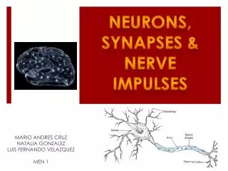
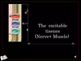
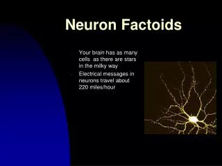
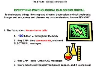
INTRODUCTION • What is a neuron? It is the basic structural and functional unit of the nervous system. It is a highly differentiatedand specialized excitable tissue. • The Human NS contains 100 billion neurons. (Nerve cells and neurons are the same.)
The nerves allow you to react to a stimulus. Stimulus A stimulus is a change in the environment. Example: A hot stove Or… tripping over a rock
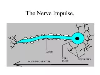
Functions of the Neurons • Reception of the stimulus • Generation of the nerve Impulse • Transmission of the nerve Impulse
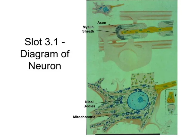
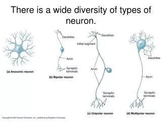
Structure of the A Typical Neuron A typical neuron thus has the following parts: • Soma or Nerve Cell body • Axon with the axon terminals • Dendrites
Different shapes Fusiform, stellate, oval, rounded, pyramidal. Different sizes – 5 to 135 micrometers Nucleus: typically large one nucleolus (usually) Perikaryon= cytoplasm which has: Nissl bodies Neurofibrils All organelles: – mitochondria, ribosomes, endoplasmic reticulum, lysosomes & Golgi apparatus SOMA (Nerve cell body)
Nissl bodies Are rough endoplasmic reticulum with ribosomes Stained with basic dyes Composed of RNA & polysomes. Tigroid substance (due to striped appearance) Not present in the axon Synthesis of proteins Dissolve & disappear if cell injured (nerve cut, injured, fatigued, poisoned) Neurofibrils Formed by clumping of neurotubules & neurofilaments Delicate threads running from cytoplasm of the nerve cell body into the axon and the dendrite Functions: Neuronal microtubules transport substances from the cell body to the distal cell processes. Neurofibrils give support and shape to the neuron. Nerve Cell Body
Point to remember: • Nerve cell body is the most vital part-if it is destroyed the entire neuron dies!
Axon • Also called axis cylinder or nerve fiber. • Longest process • A single axon arises from a cone-shaped area of the neuronal cell body called the axon hillock • Axon hillock & first 100 µm of axon (no myelin sheath) is called Initial segment. • Trigger zone: is the name given to the axon hillock & the initial segment. It is an area that shows high excitability and a nerve impulse is generated here.
AXON IS MADE UP OF: Jelly-like semi fluid substance called Axoplasm Plasma membrane called Axolemma Mitochondria and ER No Nissl granules so Does NOT synthesize proteins. AXON ENDS IN: Terminal Buttons (Synaptic knob or Bouton Terminaux) Axon break up into no. of terminal branches called Telodendria or Terminal filaments At their end is a small swelling called Terminal knob. These knobs contain granules or vesicles with neurotransmitter substance Axon
Dendrites • Short, tree-like, highly branched tapering processes of the nerve cell • Receive and then carry impulses to the cell body • Small knob-like projections called dendritic spines • Have all the components of the cell body
Afferent When nerve fiber carries impulses from the periphery towards the CNS, it is called an Afferent nerve fiber. Efferent When the nerve fiber carries impulses from the CNS to the periphery, it is called an Efferent nerve fiber. What are Afferent and Efferent fibers?
What is myelination? • Myelination is the presence of myelin around the neuron. Myelin is not part of the structure of the neuron but consists of a thick layer mostly made up of lipids, present at regular intervals along the length of the axon. • Such fibers are called myelinated fibers. • The water-soluble ions carrying the current across the membrane cannot permeate this coat, it act as an insulator, just like the white coating of the electric wires and prevents the leakage of ions from the neuron through its membrane.
How does the process of myelination occur? Myelination is carried out by myelin-forming cells that wrap themselves around the axons in jelly-roll fashion. These myelin-forming cells are Schwann cells in the PNS (peripheral nervous system) and the Oligodendrocytes in the CNS (brain & the spinal cord)
Outside CNS ↓ Schwann cells ↓ Neurons CAN regenerate ↓ Neurons can recover after injury Inside CNS ↓ Oligodendrocytes ↓ Neurons CANNOT regenerate ↓ Neurons DIE after injury Myelination
Outside the CNS: myelinated fibers • Myelination is not part of the neuron but is done by the schwann cells. • As the diagram shows, the nerve cell invaginates the schwann cell… • The schwann cell wraps around the axon in concentric spirals. • Collectively, the various layers form the myelin sheath (a patch of myelin might be made of upto 300 layers of wrapped lipid bilayers)
Nodes of Ranvier • In myelinated nerve fiber, the myelin sheath is not a continuous sheath, but is deficient at regular intervals. • Between the myelinated regions, at the NODES OF RANVIER, the axonal membrane is bare and exposed to the ECF. • Current can flow across the membrane only at these bare spaces to produce action potentials. • Voltage-gated Na+ channels are concentrated at these regions.
SALTATORY CONDUCTION In a myelinated nerve fiber, the nerve impulse “jumps” from node to node skipping over the myelinated sections of the axons. This process is called Saltatory conduction. Basis:Saltatory conduction propagates nerve impulse more rapidly because the nerve impulse has to be generated only at the nodes of ranvier and not repeatedly. Thus, it is faster. In unmyelinated fibers, the nerve impulse is like a grasshopper walking while in a myelinated fiber, the nerve impulse is like grasshopper jumping.