Download
1 / 47
510 likes | 960 Vues
Challenging Cases in Hepatology and Gastroenterology. Sanjiv Chopra, M.D., MACP Professor of Medicine Faculty Dean, Continuing Education Harvard Medical School Senior Consultant in Hepatology Beth Israel Deaconess Medical Center Boston, Massachusetts.

E N D
Challenging Cases in Hepatology and Gastroenterology Sanjiv Chopra, M.D., MACP Professor of Medicine Faculty Dean, Continuing Education Harvard Medical School Senior Consultant in Hepatology Beth Israel Deaconess Medical Center Boston, Massachusetts
We have no financial relationships I have no financial relationships with commercial entities producing, marketing, re-selling, or distributing health care goods or services consumed by, or used on, patients relevant to the content I am presenting
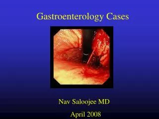
1. A 48 yr old alcoholic man is noted to have new-onset ascites; a diagnostic paracentesis yields milky fluid with a triglyceride level of 382 mg/dl. All of the following statements regarding chylous ascites are true EXCEPT: • Chylous ascites may be seen in patients with lymphoma • Chylous ascites may be seen in patients with peritoneal tuberculosis • Chylous ascites may be seen in patients with carcinoid syndrome • Chylous ascites may be seen in patients with lung cancer • Chylous may occur following abdominal trauma
Chylous Ascites and Bloody Ascites TB Tumor Trauma
Chylous Ascites and Secretory Diarrhea Carcinoid Syndrome
2. A 32 yr old woman complains of fevers, drenching night sweats, arthralgias and weakness for 2 weeks. She had a similar episode 2 years earlier. She reports that at that time she had abnormal “LFT’s” but no definitive diagnosis was made. Laboratory data from 2 years ago showed: Serum ALT 348 IU/L, AST 329 IU/L, alkaline phosphatase 392 IU/L, total bilirubin 5.8, direct bilirubin 3.9, albumin 3.0, PT 13.1. WBC 1500; 50 polys, 0 bands, 40 lymphs. Bone marrow biopsy normal. Hepatitis A, B, and C serologies negative.
Continued The patient has no prior history of surgery or no known drug allergies. She does not smoke, but has one Gin and tonic every night. Medications include oral contraceptive pills and Omeprazole. She is an RN and works in medical marketing. Physical examination is notable for a jaundiced woman in no acute distress. Her temperature is 102 F, BP 100/64 mm of Hg, pulse rate of 98 per minute. Cardiovascular, pulmonary and abdominal exam are within normal limits and she has no peripheral stigmata of chronic liver disease.
Continued Laboratory data: WBC 4000; 64 polys, 32 lymphs. Hct 33%; platelets 150,000. ALT 198 IU/L. AST 179 IU/L, alkaline phosphatase 163, total bilirubin 4.8 mg/dl, direct bilirubin 3.4 mg/dl. Albumin 2.6, PT 12.9. ANA 1:80. Blood cultures negative. Further workup: Serum copper, ceruloplasmin within normal limits. Iron studies normal. Specific autoantibodies negative; SPEP, AMA negative. Abdominal CT and chest x-ray normal. Hepatitis A, B and C serologies are negative.
Continued A percutaneous liver biopsy is performed and the results will be shown. 1. What are the common causes of this lesion? 2. What workup is indicated? 3. Are there any tantalizing clues in the patient’s history?
Granulomas • Specific inflammatory reaction • Circumscribed lesion • Central accumulation of mononuclear cells, primarily macrophages • Macrophages fuse to form multinucleated giant cells • Surrounding rim of lymphocytes, fibroblasts
Multinucleated giant cells • Fused macrophages • Secrete a variety of proteins: Lysozyme Collagenase ACE
Varieties of Granulomas • Non-caseating (eg sarcoid) • Caseating, ie central necrosis (TB) • Fibrin ring ( Q fever, HAV, Hodgkin’s, CMV,leishmaniasis, giant cell arteritis) • Lipogranulomas (mineral oil ingestion)
Disease Categories • Systemic infection • Malignancy • Drug • Autoimmune • Idiopathic
TB AIDS related MAI Crypto Fungal Histo Cocci Schistosomiasis Leprosy Brucellosis Q Fever Syphilis Cat scratch Whipple’s Infections
Malignancies • Hodgkin’s Disease • Non Hodgkin’s lumphoma • Renal Cell Carcinoma
Allopurinol Sulfonamides Chlorproprmide Quinidine Quinine Phenytoin methyldopa Carbamazepine Diltiazem Gold Hydralazine Interferon Procainamide Drugs
Miscellaneous Causes • Primary biliary cirrhosis (AMA) • Wegener’s • Giant cell arteritis • Berryliosis; talc; copper (vineyard workers) • Mineral oil ingestion • Crohn’s Disease • Idiopathic
Neat Way To Think About Granulomas • You knewthe dx PBC • You strongly suspected the dx Sarcoidosis • You see the dx Schistosomiasis TB • You don’t have the foggiest idea !
So, what is the diagnosis? • Idiopathic granulomatous hepatitis • Sarcoid • Hodgkin’s Disease • Drug
Her PMH and Social History • Meds: OCP’s, omeprazole • No prior surgery; No known drug allergies • Habits- rare cigarettes; 1 G &T nightly • Registered nurse working in medical marketing • 2 yrs earlier illness with striking similarities
Sometimes it takes a hunch… and a clever medical student!
The Diagnosis Quinine induced Granulomatous Hepatitis
We were able to get copies of her old records • She had a liver biopsy before (which she never told us) • It showed hepatic granulomas • It was 2 yrs earlier and her doctors read the same article we found and advised her NOT EVER to drink tonic or take quinine!
Her Hospital Course • She recalled that she had a biopsy after we asked again • Her fevers disappeared; white count returned to normal and her LFTs all normalized! • She left the hospital after 10 days and did not return for a scheduled f/u appointment.
Feigned Illnesses • Malingerers (external incentive such as avoiding work) • Somatization disorder (hypochondriasis, conversion reactions) • Factitious disorder
Factitious Disorders • First recognized in 2nd century AD • Most extreme form is Munchausen Syndrome
Munchausen’s Syndrome • Named after Baron Karl Friedrich von Munchausen • Can include extensive travel, multiple procedures and operations • Munchausen by Proxy (fabricating illness in a child)
Unusual Cause of Jaundice 3. A 63 year old man is referred for worsening jaundice of unclear etiology. He first noticed his eyes were yellow three weeks earlier. No past history of jaundice or liver disease. No new medicines. He does not drink any alcohol and takes no medicine other than Vitamin D3 and a daily aspirin. Family history is unremarkable. He has noted a lack of appetite and a seven pound weight loss.
At physical examination, he is clearly jaundiced but has no peripheral stigmata of chronic liver disease. There is no hepatosplenomegaly or ascites and no discernible lymphadenopathy. There are no features of portal hypertension or hepatic encephalopathy.
Laboratory Data reveal a normal CBC, PT, platelet count. His total bilirubin is 22 with a direct fraction of 15. His ALT is 68, AST 64, alkaline phosphatase 142. Serum albumin is 4.0. Hepatitis serologies are unremarkable. Iron studies are normal. ANA, smooth muscle antibody, IgG, IgM and AMA are negative or normal.
Ultrasound shows no gallstones and no biliary dilatation. A CT scan of the abdomen is normal. A liver biopsy is performed and reveals cholestasis and no definitive diagnosis. He is referred for an ERCP.
How would you define cholestasis? Should the ERCP be performed ?
A diagnostic procedure is performed. What is it ?
Chest x-ray reveals mediastinal lymphadenopathy. Biopsy of lymph nodes reveals Hodgkin’s lymphoma.
Mechanisms of Jaundice in Hodgkin’s • Mets to the porta hepatis • Massive intrahepatic metastasis • Hemolysis • Vanishing bile duct syndrome • Paraneoplastic phenomena
Jaundice can also be seen as a paraneoplastic phenomenon in patients with Hypernephroma. This is referred to as Nephrogenic Hepatic Dysfunction Syndrome or Staufer’s Syndrome.
3 Situations in Adults • Acute Cholangitis • Massive hemolysis • Fulminant hepatic failure
Causes of AFHF A HAV, Autoimmune Hepatitis B HBV C HCV D Drugs and toxins (numerous) E HEV and an Esotericdisease – Wilson’s Disease F Fatty liver (microvesicular – Pregnancy, Reye’s) G H Herpes I Iatrogenic (example chemoembolization)
What Happened to G ?! GOD only knows !