Download

1 / 18
180 likes | 311 Vues
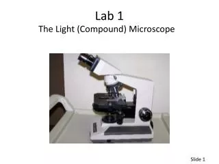
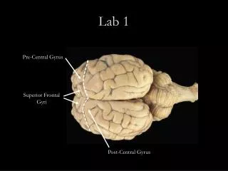
Lab 1. Planes and Sections see xv in lab manual Sagital Mid-sagital Para-sagital Frontal Transverse. Directional Terms – See xiv in lab manual Right - Left Medial – Lateral Distal – Proximal Dorsal – Ventral = Anterior – Posterior Superior – Inferior = Cranial – Caudal

E N D
Planes and Sections see xv in lab manual • Sagital • Mid-sagital • Para-sagital • Frontal • Transverse
Directional Terms – See xiv in lab manual • Right - Left • Medial – Lateral • Distal – Proximal • Dorsal – Ventral = Anterior – Posterior • Superior – Inferior = Cranial – Caudal • Superficial - Deep
Axial Cephalic Cervical Thoracic axillary Abdominal Lumbar Pelvic inguinal Appendicular Acromial Brachial Cubital Antebrachial Carpal Palmar gluteal Femoral Popliteal Crural Sural Tarsal Body Regions
Tissues • Epithelial • Connective • Muscle • Nervous
HW - 1 • Sketches of specific tissues • No more than 4 per page • Single sided • List major characteristics and where typically found • Due 6/25 beginning of lab – prior to exam • Diminished credit if turned in late
Epithelial p 56+ in text • Common characteristics • Types • Keratinized and nonkeratinized stratified squamous • Simple squamous • Simple columnar • Simple Cuboidal • Pseudostratified ciliated columnar
Epithelial Tissue Functions: protection, absorption, secretion Cover all virtually all body surfaces, in and out Lack vascular supply Classified based on shape and stacking of cells Generally supported on a basement membrane (& connective tissue)
Classification of epithelial tissues 1) Cell shape (at free surface) a) squamous - flat cells b) cuboidal - cube shaped c) columnar - column shaped Reminders: -histology = thin section of a 3D object -there are MANY tissues on each slide
Classification of epithelial tissues 2) Cell layers/arrangement a) simple - a single layer - relatively thin/fragile - found lining internal structures - simple squamous epithelium pictured here b) stratified -multiple layers -thicker -may be in areas w/ stress -stratified squamous epithelium w/out keratin pictured here
Name this epithelial tissue: Cell arrangement/layers…….Shape of cells at surface
Pseudostratified Columnar (Fig 3.6.b): -Lining much of the respiratory tract -often have cilia & goblet cells -looks layered BUT there is really only 1 layer of cells with nuclei at different heights
Name this epithelial tissue: Cell arrangement/layers…….Shape of cells at surface
Name this epithelial tissue: Figure 3.6b
Connective p 64+ in text • Common characteristics • Types • Dense • Cartilage • Bone • areolar • Adipose • Blood
Connective Tissue Most abundant tissue in body Not on free surfaces Cells separated in a matrix (ground substance + fibers) Good nerve & blood supply (except cartilage & tendons- dense connective) Classified based on matrix components, arrangement and type of protein fibers