Download
1 / 23
450 likes | 2.6k Vues
DETERMINATION OF BLOOD GLUCOSE CONCENTRATION. SITI ANNISA DEVI TRUSDA BIOCHEMISTRY DEPARTMENT. INTRODUCTION. Needed for supporting the D/ of some diseases, including DM. When to measure: fasting state or 2 hours after taking a meal.

E N D
DETERMINATION OF BLOOD GLUCOSE CONCENTRATION SITI ANNISA DEVI TRUSDA BIOCHEMISTRY DEPARTMENT
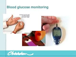
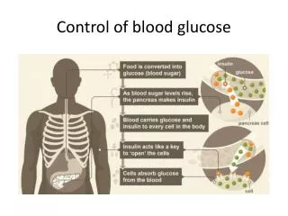
INTRODUCTION • Needed for supporting the D/ of some diseases, including DM. • When to measure: fasting state or 2 hours after taking a meal. • Sometimes at a present time detecting a present increased or decreased of blood glucose level (hyperglycemia or hypoglycemia).
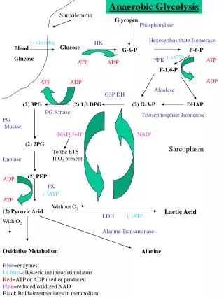
Two Methods of Measuring Blood Glucose Level • Reduction method, which is based on the ability of glucose to reduce Cu++ to Cu+less sensitive, substances that could reduce Cu++: fructose, galactose,vitaminC, creatinin, uric acid, glutathion, etc. • Enzymatic method (more specific and precise result) : Glucose is oxidized by glucose oxidase gluconicacid + H2O2 red dye.
Materials and equipments : Color reagent glucose oxidase (GOD-PAP) : • Glucose oxidase (GOD) > 15 KU/l • Peroxidase (POD) > 1.5 KU/l • 4-aminophenazone 0.25 mmol/l • Phenol 0.75 mmol/l, mutarotase > 2.0 KU/l • Phosphate buffer pH 7.5, 0.1 mol/l. Glucose standard solution 100 mg/dl (5.55 mmol/l). Serum or plasma. Aquadest. Micropipette 10 l, 1.0 ml Reaction tubes Waterbath37C Centrifuge Spectrophotometer (wavelength 492-546 nm)
To make the GOD color reagent : dissolve the content of one bottle of GOD enzyme with its solvent available. This solution is stable for 30 days at 2 – 8 oC temperature. • The absorbancy of the blank’s reagent must be about 0.0 – 0.4 AU if it is read at 505 nm wavelength compared with aquadest. • For every series of measurement, only one standard and one blank are needed.
Procedure : • 1. Centrifuge 3 ml EDTA blood, 2000 rpm for 10’. The plasma will be separated from the blood cells. Use plasma for sample • 2. Pipette into each of the three reaction tubes according to the following table :
3. Mix the content of each tube well, then incubate them at 37oC for 10 minutes or let stand at room temperature for 25 minutes. Avoid direct sunlight • 4. Using cuvette tube, read the sample’s and standard’s absorbancyagainst the blank at 505 nm.
Notes : • By this method the blood glucose level can be measured linearly up to 600 mg/dl. If the blood glucose level is higher than 600 mg/dl, dilute the plasma three times by adding 2 volumes of aquadest, then repeat the procedure. Multiply the result three times. • The result is not influenced by blood creatinin, fructose, galactose, uric acid, glutathion, ascorbic acid or bilirubin as long as these substances’s concentration are at normal range. • Bilirubin concentration up to 10 mg/dl does not influence the result, but a high dose of oral ascorbic acid (vitamin C) could decrease the result. • The color produced will be stable for 2 hours.
IDENTIFICATION OF GLUCOSE IN URINE • Urine Reduction Test (Benedict’s Test) : • Principle : • Glucose reduces the alkaline copper solution, Cu++Cu+ and is precipitated as Cu2O, a red brick color substance. • Depend on concentration of Cu2O produced, the shaked solution will produce different color that can be used to estimate the glucose concentration
Materials and equipments : Urine of a diabetic patient as sample Benedict’s qualitative solution (semi qualitative) : - 17 gr CuSO4 - 100 gr NaCO3 anhydride - 170 gr tri – Na- Citrate 2 H2O Dissolved in 1000 ml of aquadest Test tubes Waterbath100 0C/ Bunsen burner
Procedure : • Mix 3 ml Benedict’s solution with 3 drops of urine in a test tube, heat it in 100oC waterbath or just boil it for a moment, note the color produced after shaking the tube. • Repeat the same procedure with the urine diluted 2, 4 and 8 times.
Note : • The positive result of Benedict’s test could be given by many reductors, such as : • - Sugars : glucose, pentose, lactose, galactose. • - Drugs : antipyrine, pyramidon, PAS, xanthonin. • - High concentration of normally urine substances : indican, uric acid, creatinin. • - Preservative agents :formalin, CHCl3.
To read the result, shake the tube and then note the solution’s color produced : • - Blue : (-) → means there is no glucose in the sample urine • - Green : (+) → 0.5 – 1 gr % of glucose • - Yellowish green : (++) → 1 – 1.5 gr % of glucose • - Yellow : (+++) → 2 – 3.5 gr % glucose • - Red brick color : (++++) → 4 gr % of glucose