Download
1 / 1
10 likes | 155 Vues
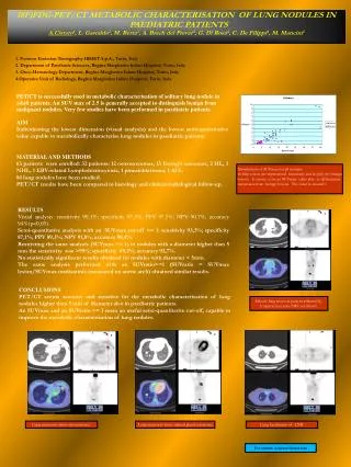
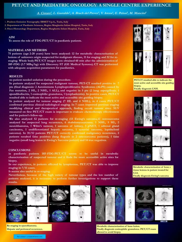
PET/CT AND PAEDIATRIC ONCOLOGY: A SINGLE CENTRE EXPERIENCE A. Cistaro 1 , L. Gastaldo 2 , A. Brach del Prever 3 , V. Arena 1 , E. Pelosi 1 , M. Mancini 1. 1 . Positron Emission Tomography IRMET S.p.A., Turin, Italy

E N D
PET/CT AND PAEDIATRIC ONCOLOGY: A SINGLE CENTRE EXPERIENCE A. Cistaro1, L. Gastaldo2, A. Brach del Prever3, V. Arena1, E. Pelosi1, M. Mancini1 1. Positron Emission Tomography IRMET S.p.A., Turin, Italy 2. Department of Paediatric Sciences, Regina Margherita Infant Hospital, Turin, Italy 3. Onco-Hematology Department, Regina Margherita Infant Hospital, Turin, Italy AIM To assess the role of FDG-PET/CT in paediatric patients. MATERIAL AND METHODS 75 patients (age2-20 years) have been analysed: 12 for metabolic characterisation of lesions of unknown origin suspected for malignant disease, 11 for staging and 52 for re-staging. Whole bodyPET/CT images were obtained 60 min after the administrationof 18F-FDG (3.7 MBq/kg) with Discovery ST (GE Medical Systems).CT was performed with adequate acquisition parameters for paediatrics. RESULTS no patient needed sedation during the procedure. In patients analysed for suspected malignant tumour, PET-CT resulted positive in 7 pts (final diagnosis: 2 Autoimmune Lymphoproliferative Syndromes (ALPS) caused by Fas mutation, 2 HL, 2 NHL, 1 ALL),and negative in 5 pts (2 lung aspergillosis, 1 spondilodiscitis, 1 eosinophilic granuloma, 1 lymphadenitis). In positive cases, PET-CT resulted able to indicate the most active and accessible site guiding biopsy. In patient analysed for tumour staging (7 HL and 4 NHL), in 4 cases PET/CT confirmed previous clinical-radiological staging. In 7 cases improved previous staging modifying clinical and therapeutical approach, finding occult tumour sites. SUV measured on first PET/CT exam is important to evaluate chemotherapy effectiveness and for patient’s follow-up. We also analysed 52 patients for re-staging (13 Ewing’s sarcoma,12 osteosarcoma analysed for suspected lung recurrence, 6 rhabdomyosarcoma, 5 NHL, 5 HL, 2 neuroblastoma, 1 Wilm’s tumour, 1 sarcoma of kidney, 3 pNET, 1 adrenal gland carcinoma, 1 undifferentiated hepatic sarcoma, 1 synovial sarcoma, 1epithelioid sarcoma). In 35/52 patients PET/CT correctly confirmed malignancy recurrence, 2 patients resulted false positives (lung flogosis in 2 osteosarcoma patients), 1 false negative (small lung lesion in Ewing’s Sarcoma patient) and 14 true negatives. PET-CT resulted able to indicate the most active and accessible site guiding biopsy. Finally diagnosis LNH. CONCLUSIONS in paediatric patients 18F-FDG-PET/CT seems to be useful in metabolic characterisation of suspected tumour and it finds the more accessible active sites for biopsy. In our experience, in patients affected by lymphomas, PET/CT was able to improve staging in 7/11 cases. It seems also useful in re-staging. Nevertheless, because of the high variety of tumour types and the low number of patients analysed, it is necessary to perform further investigations to support these results. Metabolic characterisation of bone e tissue lesions in patient treated for LLA. Finally diagnosis Ewing’s sarcoma Re-staging in epatoblastoma. Hepatic and peritoneal recurrence. Metabolic characterisation of bone lesion. Finally diagnosis eosinophilic granuloma. PET/CT exam allowed to avoid biopsy. For contact: a.cistaro@irmet.com