Download
1 / 71
740 likes | 932 Vues
hypoxia. 王婧婧 山东大学医学院病理生理学教研室. 氧气的获得和利用 :. Tissue cells. Alveolar capillary. Gas transport. air. alveoli. internal respiration. ventilation. gas exchange. external respiration. Concept. 运送氧 或 利用氧 发生障碍时,机体发生 功能 、 代谢 、 形态结构改变 的病理过程.

E N D
hypoxia 王婧婧 山东大学医学院病理生理学教研室
氧气的获得和利用: Tissue cells Alveolar capillary Gas transport air alveoli internalrespiration ventilation gasexchange externalrespiration
Concept 运送氧或利用氧发生障碍时,机体发生功能、代谢、形态结构改变的病理过程. Tissue cells can’t obtain enough oxygen or can’t fully utilize oxygen→ metabolic, functional and structural changes
Parameters of blood gas 1. Partial pressure of O2(PO2) 2. O2 capacity( CO2 max ) 3. O2 content(CO2) 4. O2 saturation( SO2)
1. Partial pressure of O2(PO2) 溶解在血液中的氧所产生的张力。 The pressure or tension produced by physically dissolved O2 in the blood. Normal: PaO2: 100 mmHg Influenced by: the oxygen pressure in the inhalied air extra-respiration 吸入气氧分压 Normal: PvO2: 40 mmHg Influenced by intra-respiration
2. O2 capacity(CO2 max) 100 ml 血液中的Hb被氧充分饱和时的最大携氧量 Maximum amount of O2 that can be combined chemically with the Hb in 100 mlblood. Normal: 20 ml/dl (15g Hb/100 ml blood ) Influenced by the amount of quality and quantity of Hb
3. O2 content(CO2) 100 ml Hb的实际携氧量 The concentration of O2 in a blood sample Normal: CaO2: 19 ml/dl,CvO2: 14 ml/dl Influenced by the PO2 and CO2 max The difference between CaO2 and CvO2 动静脉血氧差: 动脉血氧含量 - 静脉血氧含量
O2 O2 O2 O2 O2 CaO2 -- CvO2 Reflect the oxygen consumption rate of the tissue. 19ml/dl 14ml/dl A V 5ml/dl
4. O2 saturation( SO2) 指Hb结合氧的百分数 Amount of oxygen actually combined with Hb, expressed as a percentage of oxygen capacity. • CO2 – O2 of physical solution • CO2 max = 100% Normal: SaO2 95% SvO2 75% Influenced by the PO2
2,3-DPG P50 2,3-DPG
Hypoxia • Introduction • Classification, etiology and pathogenesis • Functional and metabolic changes • Factors involved in tolerance to hypoxia • Oxygen treatment and oxygen toxicity
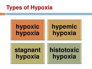
低张性缺氧 Hypotonic hypoxia 血液性缺氧 Hemic hypoxia 循环性缺氧 Circulatory hypoxia 组织性缺氧 Histogenous hypoxia Classification Gas transport +Hb circulation External respiration Tissue cells Air
循环性缺氧 Circulatory hypoxia 组织性缺氧 Histogenous hypoxia 低张性缺氧 Hypotonic hypoxia 血液性缺氧 Hemic hypoxia
1. Hypotonic hypoxia — PaO2↓ Causes : 1) PiO2↓: high altitude (3000~4000m) • 海拔高度 大气压 PiO2 PAO2 SO2 • (m) (mmHg) (mmHg) (mmHg) % • 海平面760 159 105 95 • 1000 680 140 90 94 • 2000 600 125 70 92 • 3000 530 110 62 90 • 5000 405 85 45 75 • 366 74 40 70 • 8000 270 56 30 50
100 80 60 40 20 3000 m 20 40 60 80 100 氧分压与氧饱和度的关系—— 氧离曲线 氧 饱 和 度 % pH↓ 2,3-DPG↑ Temp ↑ NO ↑ 氧分压 (mmHg)
气道狭窄 或阻塞 胸壁损伤 呼吸中枢抑制 弹性阻力增加 脊髓高 位损伤 呼吸肌 无力 脊髓前角 细胞受损 运动神经受损 2) Dysfunction of external respiration Hypoventilation
Bronchioles normal PAO2 normal PaO2 normal Bronchioles constricted PAO2 low PaO2 low Normal lung bronchial asthma
O2 CO2 表面活性物质 gas exchange disorders 血液 肺泡 内皮细胞 上皮细胞 基膜 Ventilation-perfusion mismatching Diffusion disorders
Bronchioles normal PAO2 normal PaO2 normal PAO2 normal Increased diffusion distance PaO2 low Normal lung pulmonary edema
阻塞性睡眠呼吸暂停综合征 Obstructive sleep apnea-hypopnea syndrome, OSAHS 睡时上气道塌陷阻塞引起的呼吸暂停和通气不足、伴有打鼾、睡眠结构紊乱、频繁发生血氧饱和度下降、白天嗜睡等病征。呼吸暂停是指睡眠过程中口鼻气流停止≥10s. 打鼾(打呼噜) 打鼾是睡眠期间上呼吸道气流通过时冲击咽部粘膜边缘和粘膜表面分泌物引起振动而产生的声音;其部位始至鼻咽直至下咽,包括软腭、悬雍垂、扁桃体及腭咽弓、腭舌弓、舌根、咽部的肌肉和粘膜,超过60分贝以上称为鼾症,伴有不同程度的缺氧症状时也就是睡眠呼吸暂停综合征。 治疗:减肥(超重可以引起胸壁的肥厚和腹部横膈向下运动的阻力,肥胖者的粗颈还可以增加气道的阻力)戒酒(饮酒后上呼吸道的肌肉会放松)防止平卧位(重力使舌根后坠阻塞呼吸道)使用齿具(抬高软腭,将舌根向前推,将下腭向前推)手术等。
室间隔缺损 伴肺动脉高压 右向左分流 3) Shunt 静脉血分流入动脉
①肺动脉流出道狭窄; ②室间隔膜部巨大缺损 ③主动脉右移,骑跨于室间隔缺损上方 ④右心室高度肥大及扩张 Fallot 四联症 发绀 活动时喜欢蹲踞
≥5g/dl 缺氧 发绀 (cyanosis) 毛细血管中脱氧血红蛋白≥5g/dl,使皮肤、粘膜呈青紫色. 2.6g/dl HHb HbO2 正常
紫绀与 缺氧的关系 • 缺氧不一定有发绀,发绀不一定有缺氧。 • 当血红蛋白过多或过少 时,发绀与缺氧常不一致。例如重度贫血患者,血红蛋白可降至50 g/L(5 g/dl)以下,出现严重缺氧,但不会发生紫绀。红细胞增多病患者,血中还原血红蛋白超过50 g/L (5 g/dl),出现发绀,但可无缺氧症状。
Characteristics of blood oxygen 慢性代偿 《60mmHg 发绀 慢性代偿
Hb量↓,质改变→血液携带氧的能力↓或Hb结合的氧不易释出→组织缺氧Hb量↓,质改变→血液携带氧的能力↓或Hb结合的氧不易释出→组织缺氧 Ⅱ. hemic hypoxia PaO2 SaO2正常 isotonic hypoxemia 1. Causes : (1)Hb量↓—— anemia anemic hypoxia (2)Hb质改变---- carboxyhemoglobinemia CO 中毒 1/10 a. Hb + CO Hb-CO O2 1/2100
CO O2 O2 O2 Hb: 22 b. CO抑制RBC内糖酵解→2,3-DPG生成↓→氧解离曲线左移 Hb + CO →Hb和O2的亲和力↑→释放O2↓ 樱桃红色
氧化 Hb-Fe2+ HbFe3+OH 还原 高铁血红蛋白血症(methemoglobinemia) 肠源性紫绀 (enterogenous cyanosis) 咖啡色 HbFe3+OH Fe3+不能携氧 Fe2+-O2不能解离
P50↓:Hb与O2的亲和力异常 transfusion of depot blood→2,3-DPG↓ OH-↑→pH↑ RBC内2,3-DPG含量第七天可由4.8μg/ml降至1.2μg/ml
Characteristics of blood oxygen CO, P50↓ HbFe3+OH P50↓
Color of skin anemia —— pale Hb-CO —— 樱桃红 HbFe+3-OH —— 咖啡色 无发绀 肠源性紫绀 (enterogenous cyanosis)
Ⅲ. circulatory hypoxia circulation↓ “hypokinetic hypoxia” 血液循环发生障碍,组织供血量↓ 引起的缺氧. Generalized circulatory deficiency: shock, heart failure Local circulatory deficiency: embolism, AS, thrombosis Ischemic hypoxia : Congestive hypoxia:
动脉 静脉 毛细血管内压↓ 动脉 静脉 毛细血管内压↑ • 缺血: ischemic hypoxia color of skin: pale • 淤血: Congestive hypoxia color of skin: cyanosis
O2 O2 O2 O2 O2 O2 O2 O2 19ml/dl 12ml/dl A V 7ml/dl 缺血,淤血血流缓慢血液流经cap的时间↑细胞摄氧↑CO2a-v↓ 单位时间流经组织的总血量弥散入细胞的总氧量 氧供
Inability of the cells to utilize the oxygen 组织细胞利用氧障碍 Ⅳ. histogenous hypoxia Causes: (1) “Histotoxic hypoxia” 抑制细胞氧化磷酸化: • CN-与线粒体中氧化型细胞色素氧化酶上Fe3+结合,使其不能还原,失去传递电子的功能,呼吸链中断 • 砷化物抑制细胞色素氧化酶、呼吸链酶复合物、丙酮酸氧化酶 • 甲醇通过其氧化产物甲醛与细胞色素氧化酶结合,导致呼吸链中断
(2) Mitochondria injury: endotoxin, radioactive substances, OFR (3) Vitamin deficiency: VitB1, VitPP Result : CvO2↑ →CO2a-v↓ Color of skin: HbO2↑→玫瑰色
Hb 循环障碍 Hemic hypoxia Circulatory hypoxia Hypotonic hypoxia 临床上常为混合性缺氧 失血性休克 肺淤血、水肿
Hypoxia • Introduction • Classification, etiology and pathogenesis • Functional and metabolic changes • Factors involved in tolerance to hypoxia • Oxygen treatment and oxygen toxicity Hypotonic hypoxia
1.Respiratorysystem • Compensatory response • ① PaO2< 60mmHgperipheral chemoreceptor • [H+]central chemoreceptor • alveolar ventilation↑ PaO2 ↑ PaCO2 ↓(limit) • ② Respiratory rate and depth ↑→胸内负压↑ →静脉回流↑→ 心输出量和肺血流量↑ → 有利于氧的摄取和运输
低张性缺氧引起的肺通气变化与缺氧持续的时间有关低张性缺氧引起的肺通气变化与缺氧持续的时间有关 4000m高原: 当天:通气量仅增加65%(低碳酸血症,呼碱限制) 2-3天:通气量增加5-7倍(呼碱肾代偿) 久居:通气量增加15%(外周化学感受器的敏感性↓) 肺通气量增加是急性缺氧最重要的代偿性反应 但:肺通气↑ →耗氧量↑
(2) Injured Manifestations 1)急性低张性缺氧→高原性肺水肿 快速登上4000米以上的高原,可在1~4天发生肺水肿,表现为呼吸困难、咳嗽、血性泡沫痰、肺部有湿罗音、皮肤粘膜发绀、头痛等。 男性发病率明显高于女性。缺氧、寒冷和劳累是主要诱因。轻微高原病症状如头疼、恶心及睡眠障碍等多在到达高原后的第二天至第四天出现。干咳是较早的表现,其发生可先于肺部罗音的出现。其它常见的临床表现有心动过速、低热、胸痛及白色泡沫痰甚至咯血。部分患者病情进行性恶化,出现严重的呼吸困难、精神淡漠等HAPE表现,严重者在数小时内即出现呼吸窘迫,发生急性呼吸衰竭甚至死亡。
“肺动脉高压” : 缺氧→呼吸运动↑ →胸内负压↑ 缺氧→外周血管收缩→回心血量,肺血流量↑ 缺氧性肺血管收缩→肺循环阻力↑ 局部收缩较轻或不收缩→非炎性漏出↑ 炎性因子→肺泡毛细血管膜炎性渗出↑ →肺动脉高压 cap内压↑ 肺水肿
2)中枢性呼吸衰竭 PaO2 <30mmHg 可直接抑制呼吸中枢→ 呼吸抑制,呼吸的节律和频率不规则。
2. circulatory system (1) Compensatory response ① CO ↑ → ↑氧的运输 a.心率↑(肺牵张→SN兴奋) b. 心肌收缩性↑(缺氧→ SN兴奋) c. 静脉回流量↑(呼吸 、心脏↑ ) ② pulmonary vasoconstriction“缺氧性肺血管收缩” 以调整肺内不同区域通气/血流比→ ↑氧的摄取 a.缺氧直接对VSMC作用:KV,KCa,KATP
缺氧→ KV →K+外流↓ →细胞膜去极化→电压依赖性Ca2+开放→Ca2+内流↑ → VSMC收缩→血管收缩:肺小动脉 • 胞浆游离Ca2+ ↑ →Kca开放 • ATP↓ → KATP开放 b. 体液因素:缩血管物质(AngⅡ,ET,TXA2)占优势。 c. 交感神经作用:肺血管α-肾上腺受体密度较高,交感神经兴奋时肺小动脉收缩。 K+外流↑ →血管舒张:心、脑
③ blood redistribution →保证重要器官的氧供 • 皮肤、腹腔脏器血流↓(交感兴奋,α受体密度高) • 心、脑血流↑ →(局部代谢产物,扩血管) • 心脑血管平滑肌细胞膜的KCa和KATP开放,钾外向电流增加,细胞膜超极化,Ca2+内流↓ 。 ④ cajpillary proliferation 长期缺氧 → 缺氧诱导因子-1(HIF-1) →血管内皮生长因子(VEGF)基因表达↑,尤其是心、脑、骨骼肌→毛细血管增生 → ↓氧弥散距离,↑供氧量