Download
1 / 15
200 likes | 582 Vues
Bacterial Cell Wall Hydrolysis by Lysozyme. We have examined the structure of the well-characterized hen egg white (HEW) lysozyme in considerable detail. This enzyme catalyzes the hydrolysis of bacterial wall polysaccharides of the following structure:

E N D
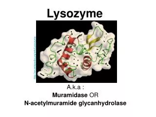
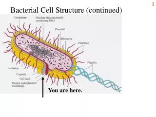
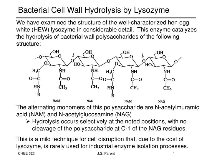
Bacterial Cell Wall Hydrolysis by Lysozyme • We have examined the structure of the well-characterized hen egg white (HEW) lysozyme in considerable detail. This enzyme catalyzes the hydrolysis of bacterial wall polysaccharides of the following structure: • The alternating monomers of this polysaccharide are N-acetylmuramic acid (NAM) and N-acetylglucosamine (NAG) • Hydrolysis occurs selectively at the noted positions, with no cleavage of the polysaccharide at C-1 of the NAG residues. • This is a mild technique for cell disruption that, due to the cost of lysozyme, is rarely used for industrial enzyme isolation processes. J.S. Parent
Characterization of the Active Site • While the mechanism of every catalytic process is very difficult to determine, enzyme mediated reactions are particularly troublesome, as we have very little knowledge of which functional groups participate directly in the catalysis. • Kinetic data is useful for design purposes, but it rarely leads to a reliable reaction mechanism without supporting information. • Product distribution analysis of model compounds that approximate the reactive functionality of the polysaccharide. • Crystallographic analysis of the enzyme and various enzyme-inhibitor or enzyme-substrate complexes • Assessment of the influence of select amino acid substituent modification • Measurement of kinetic isotope effects • All four of these techniques have been applied to lysozyme catalysis, and we will examine the first three to better understand the nature of enzyme-substrate interactions. J.S. Parent
Lysozyme Activity Studies - Model Compounds • The cleavage patterns for acetylglucosamine oligomers • are not consistent with a random attack of the enzyme. • Cleavage occurs at an appreciable rate only for hexamers or higher oligomers and occurs between mers 4 and 5. • Indicates that a hexasaccharide is incorporated by the catalytic site of the enzyme, and a unique mode of substrate activation must establish a preferred hydrolysis pathway. J.S. Parent
Crystallographic Studies of Lysozyme-Inhibitor Complexes • Once the crystal structure of an enzyme has been determined the structures of isomorphous crystals that contain additional molecules may be determined without difficulty. • This method has been used to explore the interactions between lysozyme and a wide range of substrate-related inhibitor molecules. Perspective drawing of the main chain conformation of lysozyme: Elevation from active-site side of the molecule. Only a positions of a-carbon atoms are shown. J.S. Parent
Crystallographic Studies of Lysozyme-Inhibitor Complexes • The trisaccharide of NAG forms a relatively stable complex with lysozyme that has been characterized by crystallography. The position of the three NAG groups is illustrated at the top-right of the structure. • The other three groups have been • positioned by molecular • modeling, as they cannot be • isolated. • N-acetylglucosamine (NAG) alone • binds to the enzyme with H-bonds between the NH and carbonyl • oxygens of its acetamido side chain and the main peptide chain and CO and NH groups of residues 107 and 59. J.S. Parent
Active Site Determination: Chemical Modification • If the chemical modification of a particular amino acid side chain results in enzyme deactivation, then the residue in question is located at the active site, provided that the modification can be prevented by the presence of excess substrate or inhibitor. The following is a summary of specific modifications used to determine the activity of individual residues: • A. Amino (Lysyl e-amino, 1,13, 33, 96, 97,116) • All 6 lysine residues are on the surface of the enzyme, with Lys 33 situated in the very bottom of the cleft. • Little effect of modifications of this type on lytic activity is observed, suggesting that these amino acid residues do not participate directly in catalysis. J.S. Parent
Active Site Determination: Chemical Modification • B. Arginine (5, 14, 21, 45, 61, 68, 73, 112, 114, 125, 128) • All but one Arg is located on the surface of the enzyme, but Arg 114 is believed to form two hydrogen bonds with the saccharide. • Modification of 7 of the 11 Arg residues had little influence on the activity of lysozyme on NAG4. • C. Glutamic Acid 7, 35; Aspartic Acid 18, 48, 52, 66, 87, 101, 111 • Modification studies of carboxyl groups have provided unequivocal evidence for the involvement of these residues in catalysis. • Exhaustive esterification of lysozyme with acid alcohol results in a loss of enzyme activity: J.S. Parent
Active Site Determination: Chemical Modification • C. Carboxyl Groups Continued • Two amino acid residues in particular (Asp 52 and Glu 35) have been implicated in several modification studies. • Modification of Asp 52 was inhibited by the presence of substrate • Selective oxidation of Glu 35 with iodine denatures the enzyme. • D. Cysteine (6-127, 30-115, 64-80, 76-94) • Reduction of disulfide crosslinks denatures the enzyme, although 6-127 can be opened without deactivation. This is reversible, as air oxidation regenerates enzymatic activity with high yield. • E. Histidine (15) • Alkylation of the single histidine has been shown to have little influence on lytic activity. J.S. Parent
Active Site Determination: Chemical Modification • F. Methionine (12,105) • Cyanogen bromide in 70% formic acid cleaves both methionyl peptide bonds without modification of other amino acid sequences. • This reduces activity to 10% of the native enzyme, despite the fact that both residues are buried within the enzyme structure and participate through non-polar contacts with other residues. • G. Tryptophan (28, 62, 63, 108, 111, 123) • Three of six tryptophans are believed to be positioned in the active site. Oxidation with N-bromosuccinimide inactivates the enzyme. • Selective oxidation of Trp 108 • with iodine is blocked by • substrate. Trp is also in close • proximity to Glu 35. J.S. Parent
Binding of Lysozyme to Hexa-N-Acetylglucosamine • Schematic illustration of the active site in the cleft region of lysozyme. A through F represent the glycosyl moieties of a • hexa-saccharide. Some of the amino • acids in the cleft region near • these subsites of the • active site • are shown. J.S. Parent
Model of the HEW Lysozyme Site • Schematic diagram showing the specificity of lysozyme for hexa-saccharide substrates. • Six subsites A-F on the enzyme bind the sugar residues. Alternate sites interact with the acetamido side chains (a), and these sites are unable to accommodate MurNAe residues with their lactyl side chains (P). • Site D cannot bind a sugar residue without distortion, and the glycosidic linkage that is cleaved binds between sites D and E as shown by the arrow. J.S. Parent
Proposed Catalytic Mechanism • 1. The saccharide binds in the enzyme cleft with residue D distorted to a conformation resembling the half-chair. • 2. Bond rearrangement to yield a carbenium ion proceeds at a rate enhanced through several contributions: • A. Glu 35 acts as a general acid catalyst, donating H to the glycosidic oxygen. • B. Asp 52 bears a negative charge that favors formation of the carbenium ion. • C. The ring conformation is close to that required in the transition state. • D. The nonpolar nature of the cleft possibly enhances reaction rate. • 3. The enzyme-bound carbonium ion is stabilized by neighboring charges of Asp 52 and Glu 35, the latter having deprotonated in bond rearrangement. • 4. The aglycone diffuses away, and reaction with water or another acceptor completes the process. J.S. Parent
pH Dependence of Enzyme Activity • Since the characteristics of ionizable side chains of amino acids depend on pH, enzyme activity varies with pH shifts. • At extremes of pH, the tertiary structure of the protein may be disrupted and the enzyme denatured. • Even at moderate pH values where tertiary structure is unaffected, enzyme activity may depend on the degree of ionization of certain amino acid side chains • pH can therefore affect enzyme • conformation, substrate binding • and the ability of “active” side • groups to participate in catalysis, • as shown here for three • representative enzymes. J.S. Parent
Survey of Ionizable Enzyme Groups • The ionizable groups which contribute to the acid-base properties of proteins, shown with their approximate pKa values. These can vary by several pH units depending on their environment in the protein. J.S. Parent
pH Dependence of Enzyme Activity • Recall our discussion of lysozyme, where Asp52 was believed to exist in its conjugate base (RCOO-) form, while Glu35 is thought to be active in its acidic state (RCOOH). • pH will dictate the degree of protonation of these residues, creating an optimum that is dependent on their pKa’s. • Graphs of Vmax against pH, at constant [E], where catalytic activity depends on the simultaneous presence of EY- and EZH • (a) where pKy and pKz are more than 2 units apart • (b) where pKy and pKz are less than 2 units apart J.S. Parent