Download
1 / 44
440 likes | 562 Vues
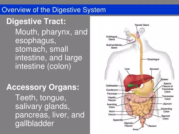
Overview of the Digestive System. Digestive Tract: Mouth, pharynx, and esophagus, stomach, small intestine, and large intestine (colon) Accessory Organs: Teeth, tongue, salivary glands, pancreas, liver, and gallbladder. Components of the Digestive System. GI Tract: Oral Cavity Pharynx

E N D
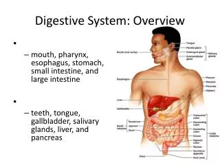
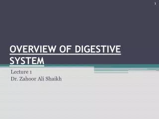
Overview of the Digestive System Digestive Tract: Mouth, pharynx, and esophagus, stomach, small intestine, and large intestine (colon) Accessory Organs: Teeth, tongue, salivary glands, pancreas, liver, and gallbladder
Components of the Digestive System • GI Tract: • Oral Cavity • Pharynx • Esophagus • Stomach • Small Intestine • Large Intestine
The Pharynx • Nasopharynx • Oropharynx • Laryngopharynx
Peristalsis in Esophagus LE 21-8 Muscles contract Muscles contract, constricting passageway and pushing bolus down Muscles relax Bolus of food Muscles relax, allowing passageway to open Muscles contract Muscles relax Stomach
Stomach • Site where food is churned into chyme • Protein digestion begins
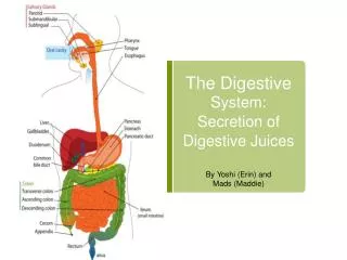
LE 21-11a Duodenum and Related Organs Liver Bile Gall- bladder Stomach Bile Pancreas Acid chyme Intestinal enzymes Pancreatic juice Duodenum of small intestine
Small Intestine – Gross Anatomy • Longest portion of the alimentary canal • Site of most enzymatic digestion and absorption • Three subdivisions • Duodenum, jejunum, and ileum
Gross Anatomy of Large Intestine • Cecum • Appendix • Ascending • Transverse • Descending • Sigmoid colon • Rectum • Anus
LE 21-12 Large intestine (colon) Small intestine Sphincter End of small intestine Rectum Anus Nutrient flow Appendix Cecum
Gross Anatomy of Large Intestine • Rectum – descends along the inferior half of the sacrum • Anal Canal – the last subdivision of the large intestine
Gallbladder • Stores and concentrates bile • Expels bile into duodenum • Bile emulsifies fats
Histology of the Digestive Tract Mucosa Submucosa Muscularis Externa Serosa
Mucosa (Innermost Layer) • Epithelium • Connective Tissue (Blood vessels, lymphatic vessels, lymphatic nodules) • Smooth Muscle
Submucosa Connective tissue • Blood vessels • Lymphatic vessels • Nerve plexus • May have glands and lymphatic tissue
Muscularis Externa Smooth or Skeletal Muscle If smooth muscle, usually 2 layers (circular and longitudinal)
Adventitia or Serosa Adventitia (organs superior to diaphragm) Serosa = visceral peritoneum
The Esophagus • Stratified squamous epithelium • Mucous glands • Muscularis externa – skeletal muscle first third of length
LE 21-11b Lumen of intestine Nutrient absorption Vein with blood en route to the liver Nutrient absorption into epithelial cells Microvilli Epithelial cells Amino acids and sugars Fatty acids and glycerol Lumen Muscle layers Fats Blood capillaries Large circular folds Blood Villi Lymph vessel Lymph Nutrient absorption Epithelial cells Villi Intestinal wall
Small Intestine: Duodenum Br = Brunner glands V = Villus G = Goblet cells Cr = Intestinal glands MM = Muscularis Mucosae LP = Lamina Propria
Microscopic Anatomy of Large Intestine • Villi are absent • Contains numerous goblet cells • Intestinal crypts – simple tubular glands • Lined with simple columnar epithelial tissue • Epithelium changes at anal canal • Becomes stratified squamous epithelium
Liver • Largest gland in the body • Performs over 500 functions • Digestive function – bile production • Performs many metabolic functions
The Peritoneal Cavity and Peritoneum • Mesentery – a double layer of peritoneum • Holds organs in place • Sites of fat storage • Provides a route for circulatory vessels and nerves
Mesenteries • Superficial view of the abdominal organs
Mesenteries • Greater omentum and transverse colon reflected
Mesenteries • Sagittal section through the abdominopelvic cavity