Download
1 / 34
370 likes | 586 Vues
The Eyes and Ears. Chapter 11 Special Senses. The Eye. The Eye. The eye is a globe shaped, hollow structure set within a bony cavity The bony cavity or orbit houses the eyeball and assorted structures Eye muscles Nerves Blood vessels

E N D
The Eyes and Ears Chapter 11Special Senses
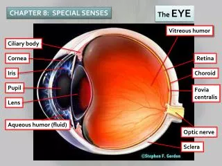
The Eye • The eye is a globe shaped, hollow structure set within a bony cavity • The bony cavity or orbit houses the eyeball and assorted structures • Eye muscles • Nerves • Blood vessels • Most of the eyeball is protected from trauma by the orbit’s bony cavity
The wall of the eye • The wall of the eye is comprised of three layers • Sclera- white outer layer of the eyeball is composed of fibrous connective tissue • Cornea- is a transparent domed structure formed by the sclera • the cornea also protects the front part of the eye from injury • Is the first part of the eye that refracts light rays. • The cornea is avascular (without blood vessels or capillaries) • Choroid- lies below the sclera • Contains blood vessels and dark pigmented tissue that prevents glare within the eyeball by absorbing light.
Other structures of the eye • Ciliary Body- is the anterior portion of the choroid • Iris- the colored portion of the eye • Retina- lines the posterior two thirds portion of the eyeball and contains rods and cones • The sensory receptors for vision • Rod- a rod-shaped receptor in the retina of the eye that is sensitive to dim light but not color • Cone- a cone-shaped cell sensitive to light and color in the retina of the eye of a human being or any other vertebrate animal. There are three different types of cone cells, responding to blue, green, or red light.
Structures continued • Pupil- the opening in the center of the iris • The amount of light entering the eye is controlled by contractions and dilations of the pupil • Optic disk- the retinal nerve fibers unite here • The optic disk is known as the “blind spot” because the optic disk has no rods or cones • Optic nerve- formed from the retinal fibers and cross through the wall of the eyeball
Vision Terms • Myopia • Nearsightedness • Unable to see clearly objects that are far away • Hyperopia • Farsightedness • Able to see distant objects better than nearby ones • Erythropia • Objects that are not red appear red • Xanthopia • Objects that are not yellow appear as yellow • Diplopia • Double vision • Emmetropia • Normal vision • Astigmatism • A defect in a lens that prevents light rays from meeting at a single point producing an imperfect image (blurred vision)
Vision Defects Emmetropia Hypertropia Myopia Astigmatism
Other structures in the Eye Socket • 1. Lacrimal Gland • Is located above the outer corner or each eye • These glands produce tears, which keep the eyeballs moist • 2. Lacrimal Sac • Collects tears • 3. Nasolacrimal ducts • Tears are drained to the nose thru the these ducts then expelled from the body thru the nose. 1 2 3
Combining forms Blephar/o eyelid blepharospasm Choroid/o choroid choroidopathy Corne/o cornea corneitis Cor/o pupil anisocoria Dacry/o tear dacryorrhea Lacrim/o tear lacrimation Dipl/o double diplopia
Combining forms • Irid/o iris iridoplegia • Kerat/o horny tissue, hard, cornea • keratoplasty • Ocul/o eye intraocular • Ophthalm/o eye ophthalm/o/scope • Opt/o eye,vision optic • Retin/o retina retinopathy • Scler/o hardening,sclera scler/itis
Suffixes -opia vision amblyopia -opsia vision heteropsia -ptosisprolapse, downward displacement blepharoptosis
Pathology of the Eye Achromatopsia- color blindness, more common in men Astigmatism- blurred vision due to defect in the curvature of the cornea and lens Cataract- opacity (cloudiness) of the lens as result of protein deposits on its surface that slowly builds up until vision is lost Conjunctivitis- inflammation of the conjunctiva that can be caused by bacteria, allergy, irritation, or a foreign body; also called “pinkeye” Diabetic retinopathy- retinal damage marked by aneurismal dilation of blood vessels
Esotropia- strabismus in which there is deviation of the visual axis of one eye toward that of the other eye resulting in diplopia (double vision) also called “cross eyed” Glaucoma- increased intraocular pressure caused by the failure of the aqueous humor (the transparent fluid that circulates in the eye chamber) to drain, which results in atrophy of the optic nerve and eventually may lead to blindness. Strabismus- muscular eye disorder in which the eyes turn from the normal position so that they deviate in different directions
Hordeolum- small purulent (relating to, containing, or consisting of pus) inflammatory infection of the sebaceous gland of the eyelid; also called a “sty” Macular degeneration- breakdown of the tissues in the macula resulting in loss of central vision Photophobia- unusual intolerance and insensitivity to light; occurs in diseases such as measles, meningitis, inflammation of the eyes, measles and rubella Retinal detachment- separation of the retina from the choroid, which disrupts vision and results in blindness if not treated.
Diagnostics Tonometry- measuring of the intraocular pressure by determining the resistance of the eyeball to indention by an applied force; used to detect glaucoma Visual acuity test- standard test of visual acuity using the “ E-Chart”
Therapeutic Cataract surgery- excision of cataracts by surgical removal of the lens Corneal transplant- (keratoplasty) surgical transplantation of a donor cornea (from a cadaver) into the eye of a recipient Extracapsular surgery- excision of most of the lens, followed by insertion of an intraocular lens transplant Phacoemulsification- excision of the lens by ultrasonic vibrations that break the lens into tiny pieces which are then suctioned out of the eye.
The Ears • The ears and their accessory structures are the receptor organs that enable us to hear and maintain our balance.
Each ear consists of three divisions • The external ear, the middle ear, and the inner ear • The External and Middle ear • Conducts sound waves through the ear • The Inner ear • Contains auditory structures that receive the sound waves and transmits them to the brain for interpretation. • Contains specialized receptors that maintain balance and equilibrium regardless of changes in the body position or motion
The External Ear • the outside part of the ear, consisting of the auricle and auditory canal • Auricle-the part of the external ear that projects outward from the head ***WARNING*** do not confuse with the auricle in the heart-an ear-shaped muscular part that sticks out from the surface of each upper chamber atrium of the heart. • Auditory Canal- a passage from the outer ear to the ear drum along which sound waves travel
The Middle Ear • The narrow air-filled space between the ear drum and the outer wall of the inner ear containing the three tiny bones that transmit sound vibrations • 1. Incus-a small anvil-shaped bone in the middle ear of mammals • 2. Stapes-a small stirrup-shaped bone in the middle ear of mammals, the innermost of the three small bones that transmit vibration to the inner ear • 3. Malleus- a hammer-shaped bone, the outermost of three small bones in the middle ear that transmit sound waves from the eardrum to the inner ear.
Bones of the Middle Ear • 1. Incus • 2. Stapes • 3. Malleous
The Inner Ear • The fluid-filled part of the ear, including the cochlea, which is responsible for hearing, and the semicircular canals, which control balance • Cochlea- a spiral structure in the inner ear that looks like a snail shell and contains tiny hair cells whose movement is interpreted by the brain as sound • Semicircular canals- each of three tubes in the inner ear, semicircular in shape and set at right angles to one another, that help to maintain balance
Eardrum • A membrane of thin skin and fibrous tissue that vibrates in response to sound waves, located between the external and the middle ear.
Pathology of the Ear • Acoustic neuroma- a benign tumor that develops on the auditory nerve causing hearing loss, loss of balance, and headaches • Anacusis-total deafness; complete hearing loss • Conductive hearing lose- hearing loss due to an impairment in the transmission of sound because of an obstruction of the ear canal or damage to the eardrum or ossicles (a small bone,especially one of three bones of the middle ear in humans) • Meniere disease- rare disorder of unknown etiology within the labyrinth (structure consisting of connected cavities or canal)
Pathology continued • Otitis media- a painful inflammation of the middle ear that can cause dizziness and temporary hearing loss • Otosclerosis- a hereditary disease of the inner ear in which spongy bone growth leads to progressive hearing impairment • Presbycusis- impairment of hearing resulting from the aging process • Tinnitus- a continual noise in the ear, e.g. a ringing or roaring, usually caused by damage to the hair cells of the inner ear • Vertigo- a condition in which somebody feels a sensation of whirling or tilting that causes a loss of balance
Diagnostic • Audiometry- test that measures hearing acuity of various sound frequencies • Otoscopy- visual examination of the ear; especially the eardrum using an otoscope • Rinne test- hearing acuity test that is performed with a vibrating tuning fork placed on the mastoid process, then in front of the external auditory canal to test bone and air conduction
Therapeutic • Cochlear implant- electronic transmitter that is surgically implanted into the cochlea of a deaf person; performed to restore hearing loss • Myringoplasty- surgical repair of a perforated eardrum with a tissue graft • Myringotomy-incision of the eardrum to relieve pressure and release pus or serous fluid from the middle ear or to insert tympanostomy tubes surgically in the eardrum
Combining forms • Acous/o hearing • Audi/o hearing • Myring/o tympanic membrane • Tympan/o tympanic membrane • Ot/o ear • Salping/o tube (usually fallopian or eustachian) auditory tube
Suffixes • -acusis hearing • -tropia turning