Download
1 / 75
800 likes | 1.21k Vues
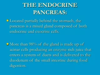
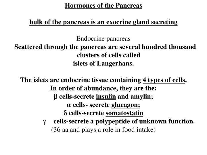
Hormones of the Pancreas bulk of the pancreas is an exocrine gland secreting Endocrine pancreas Scattered through the pancreas are several hundred thousand clusters of cells called islets of Langerhans. The islets are endocrine tissue containing 4 types of cells .

E N D
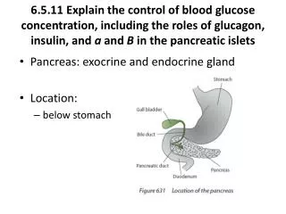
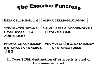
Hormones of the Pancreas • bulk of the pancreas is an exocrine gland secreting • Endocrine pancreas • Scattered through the pancreas are several hundred thousand clusters of cells called • islets of Langerhans. • The islets are endocrine tissue containing 4 types of cells. • In order of abundance, they are the: • b cells-secrete insulin and amylin; • a cells- secrete glucagon; • d cells-secrete somatostatin • cells-secrete a polypeptide of unknown function. (36 aa and plays a role in food intake)
The endocrine portion of the pancreas takes the form of many small clusters of cells called islets of Langerhans or, more simply, islets. Humans have roughly one million islets. In standard histological sections of the pancreas, islets are seen as relatively pale-staining groups of cells embedded in a sea of darker-staining exocrine tissue. The image to the right shows 3 islets in a horse pancreas.
Interestingly, the different cell types within an islet are not randomly distributed – beta cells occupy the central portion of the islet and are surrounded by a "rind" of a and d cells. Aside from the insulin, glucagon and somatostatin, a number of other "minor" hormones have been identified as products of pancreatic islets cells.
Islets are richly vascularized, allowing their secreted hormones ready access to the circulation. Although islets comprise only 1-2% of the mass of the pancreas, they receive about 10 to 15% of the pancreatic blood flow. Additionally, they are innervated by parasympathetic and sympathetic neurons, and nervous signals clearly modulate secretion of insulin and glucagon.
Insulin Synthesis and Secretion Structure of Insulin Insulin is a rather small protein, with a molecular weight of about 6000 Daltons. composed of 2 chains held together by disulfide bonds. The figure shows a molecular model of bovine insulin, with the A chain colored blue and the larger B chain green. The amino acid sequence is highly conserved among vertebrates, and insulin from one mammal almost certainly is biologically active in another. For years diabetic patients were treated with insulin extracted from pig or cow pancreases.
Biosynthesis of Insulin Insulin is synthesized in significant quantities only in b cells in the pancreas. The insulin mRNA is translated as a single chain precursor called preproinsulin, and removal of its signal peptide during insertion into the endoplasmic reticulum generates proinsulin. Proinsulin consists of three domains: an amino-terminal B chain, a carboxy-terminal A chain and a connecting peptide in the middle known as the C peptide. Within the endoplasmic reticulum, proinsulin is exposed to several specific endopeptidases which excise the C peptide, thereby generating the mature form of insulin. Insulin and free C peptide are packaged in the Golgi into secretory granules which accumulate in the cytoplasm.
Since insulin was discovered in 1921, it has become one of the most thoroughly studied molecules in scientific history.
Control of Insulin Secretion Insulin is secreted in primarily in response to elevated blood concentrations of glucose. This makes sense because insulin is "in charge" of facilitating glucose entry into cells. Some neural stimuli (e.g. site and taste of food) and increased blood concentrations of other fuel molecules, including amino acids and fatty acids, also WEAKLY promote insulin secretion. Our understanding of the mechanisms behind insulin secretion remain somewhat fragmentary. Nonetheless, certain features of this process have been clearly and repeatedly demonstrated, yielding the following model:
Control of Insulin Secretion Glucose is transported into the b cell by facilitated diffusion through a glucose transporter; elevated concentrations of glucose in extracellular fluid lead to elevated concentrations of glucose within the b cell.Elevated concentrations of glucose within the b cellultimately leads to membrane depolarization and an influx of extracellular calcium. The resulting increase in intracellular calcium is thought to be one of the primary triggers for exocytosis of insulin-containing secretory granules.
Control of Insulin Secretion The mechanisms by which elevated glucose levels within the b cell cause depolarization is not clearly established, but seems to result from metabolism of glucose and other fuel molecules within the cell, perhaps sensed as an alteration of ATP:ADP ratio and transduced into alterations in membrane conductance. Increased levels of glucose within b cells also appears to activate calcium-independent pathways that participate in insulin secretion.
Control of Insulin Secretion Stimulation of insulin release is readily observed in whole animals or people. The normal fasting blood glucose concentration in humans and most mammals is 80-90 mg per 100 ml, associated with very low levels of insulin secretion.
Control of Insulin Secretion The figure depicts the effects on insulin secretion when enough glucose is infused to maintain blood levels 2-3 times the fasting level for an hour. Almost immediately after the infusion begins, plasma insulin levels increase dramatically. This initial increase is due to secretion of preformed insulin, which is soon significantly depleted. The secondary rise in insulin reflects the considerable amount of newly synthesized insulin that is released immediately. Clearly, elevated glucose not only simulates insulin secretion, but also transcription of the insulin gene and translation of its mRNA.
Physiologic Effects of Insulin Stand on a streetcorner and ask people if they know what insulin is, and many will reply, "Doesn't it have something to do with blood sugar?" Indeed, that is correct, but such a response is a bit like saying "Mozart? Wasn't he some kind of a musician?" Insulin is a key player in the control of intermediary metabolism. It has profound effects on both carbohydrate and lipid metabolism, and significant influences on protein and mineral metabolism. Consequently, derangements in insulin signalling have widespread and devastating effects on many organs and tissues.
Physiologic Effects of Insulin The Insulin Receptor (IR) and Mechanism of Action Like the receptors for other protein hormones, the receptor for insulin is embedded in the PM The IR is composed of 2 alpha subunits and 2 beta subunits linked by S-S bonds. The alpha chains are entirely extracellular and house insulin binding domains, while the linked beta chains penetrate through the PM.
The IR is a tyrosine kinase. it functions as an enzyme that transfers phosphate groups from ATP to tyrosine residues on target proteins. Binding of insulin to the alpha subunits causes the beta subunits to phosphorylate themselves (autophosphorylation), thus activating the catalytic activity of the receptor. The activated receptor then phosphorylates a number of intracellular proteins, which in turn alters their activity, thereby generating a biological response.
Physiologic Effects of Insulin Several intracellular proteins have been identified as phosphorylation substrates for the insulin receptor, the best-studied of which is Insulin receptor substrate 1 or IRS-1. When IRS-1 is activated by phosphorylation, a lot of things happen. Among other things, IRS-1 serves as a type of docking center for recruitment and activation of other enzymes that ultimately mediate insulin's effects.
Physiologic Effects of Insulin Insulin and Carbohydrate Metabolism Glucose is liberated from dietary carbohydrate such as starch or sucrose by hydrolysis within the SI, and is then absorbed into the blood. Elevated concentrations of glucose in blood stimulate release of insulin, and insulin acts on cells thoughout the body to stimulate uptake, utilization and storage of glucose.
Physiologic Effects of Insulin Two important effects are: Insulin facilitates entry of glucose into muscle, adipose and several other tissues. The only mechanism by which cells can take up glucose is by facilitated diffusion through a family of glucose transporters. LARGELY FAT and SKELETAL MUSCLE
Physiologic Effects of Insulin Two important effects are: In many tissues - muscle being a prime example - the major transporter used for uptake of glucose (called GLUT4) is made available in the plasma membrane through the action of insulin.In the absense of insulin, GLUT4 glucose transporters are present in cytoplasmic vesicles, where they are useless for transporting glucose. Binding of insulin to IR on such cells leads rapidly to fusion of those vesicles with the plasma membrane and insertion of the glucose transporters, thereby giving the cell an ability to efficiently take up glucose. When blood levels of insulin decrease and insulin receptors are no longer occupied, the glucose transporters are recycled back into the cytoplasm.
Family of Glucose transport proteins Uniporters-transfer one molecule at a time Facillitated diffusion Energy indepednent GLUT1- found on PM every single cell in your body for glucose uptake GLUT2-liver transporter, also found in b cells GLUT3- fetal transporter GLUT4- insulin sensitive glucose transporter GLUT5- GLUT7 NOT to be confused with Na+glucose transporter in lumen of SI which is a symporter, couple the movement of glucose (against) with Na+ (with gradient)
GLUT1-glucose transporter on the plasma membrane of every cell in your body
Glucose = GLUT1 Glucose Glucose Glucose Cytoplasm Glucose Nucleus
GLUT4-a tissue specific insulin sensitive glucose transporter
Glucose = GLUT1 Glucose = GLUT4 Glucose Glucose Glucose Glucose Glucose Fat and Skeletal Muscle Cells have GLUT4 Nucleus
= GLUT1 Glucose INSULIN = GLUT4 Glucose Insulin binds its cell surface receptor Glucose GLUT4 vesicles travel to PM Nucleus
= GLUT1 Glucose INSULIN = GLUT4 Glucose Glucose Glucose Glucose Glucose Lots of glucose inside cell Nucleus
Very important that glucose is in cells and not in blood Hyperglycemia-high blood glucose
In the absense of insulin, GLUT4 glucose transporters are present in cytoplasmic vesicles, where they are useless for transporting glucose. Binding of insulin to receptors on such cells leads rapidly to fusion of those vesicles with the plasma membrane and insertion of the glucose transporters, thereby giving the cell an ability to efficiently take up glucose. When blood levels of insulin decrease and insulin receptors are no longer occupied, the glucose transporters are recycled back into the cytoplasm.
INSULIN TALK TO LIVER TO SUPPRESS HGO Hepatic glucose output GLUT2 is the liver transporter Insulin stimulates the liver to store glucose in the form of glycogen. Some glucose absorbed from the SI is immediately taken up by hepatocytes, which convert it into the storage polymer glycogen.
Insulin has several effects in liver which stimulate glycogen synthesis. First, it activates the enzyme hexokinase, which phosphorylates glucose, trapping it within the cell. Coincidently, insulin acts to inhibit the activity of glucose-6-phosphatase. Insulin also activates several of the enzymes that are directly involved in glycogen synthesis, including phosphofructokinase and glycogen synthase. The net effect is clear: when the supply of glucose is abundant, insulin "tells" the liver to bank as much of it as possible for use later.
well-known effect of insulin is to decrease the concentration of glucose in blood Another important consideration is that, as blood glucose concentrations fall, insulin secretion ceases. In the absense of insulin, a bulk of the cells in the body become unable to take up glucose, and begin a switch to using alternative fuels like fatty acids for energy. Neurons, however, require a constant supply of glucose, which in the short term, is provided from glycogen reserves. In the absense of insulin, glycogen synthesis in the liver ceases and enzymes responsible for breakdown of glycogen become active. Glycogen breakdown is stimulated not only by the absense of insulin but by the presence of glucagon which is secreted when blood glucose levels fall below the normal range.
Insulin and Lipid Metabolism The metabolic pathways for utilization of fats and carbohydrates are deeply and intricately intertwined. Considering insulin's profound effects on carbohydrate metabolism, it stands to reason that insulin also has important effects on lipid metabolism.
Insulin and Lipid Metabolism Notable effects of insulin on lipid metabolism include the following: Insulin promotes synthesis of fatty acids in the liver. As discussed above, insulin is stimulatory to synthesis of glycogen in the liver. However, as glycogen accumulates to high levels (roughly 5% of liver mass), further synthesis is strongly suppressed.When the liver is saturated with glycogen, any additional glucose taken up by hepatocytes is shunted into pathways leading to synthesis of fatty acids, which are exported from the liver as lipoproteins. The lipoproteins are ripped apart in the circulation, providing free fatty acids for use in other tissues, including adipocytes, which use them to synthesize triglyceride.
Insulin and Lipid Metabolism Insulin promotes synthesis of fatty acids in the liver. When the liver is saturated with glycogen, any additional glucose taken up by hepatocytes is shunted into pathways leading to synthesis of fatty acids, which are exported from the liver as lipoproteins. The lipoproteins are ripped apart in the circulation, providing free fatty acids for use in other tissues, including adipocytes, which use them to synthesize triglyceride.
Insulin and Lipid Metabolism Insulin inhibits breakdown of fat in adipose tissue by inhibiting the intracellular lipase that hydrolyzes triglycerides to release fatty acids.Insulin facilitates entry of glucose into adipocytes, and within those cells, glucose can be used to synthesize glycerol. This glycerol, along with the fatty acids delivered from the liver, are used to synthesize triglyceride within the adipocyte. By these mechanisms, insulin is involved in further accumulation of triglyceride in fat cells.
INSULIN IN AN ANABOLIC HORMONE From a whole body perspective, insulin has a fat-sparing effect. Not only does it drive most cells to preferentially oxidize carbohydrates instead of fatty acids for energy, insulin indirectly stimulates accumulation of fat is adipose tissue.
Other Notable Effects of Insulin (I) In addition to insulin's effect on entry of glucose into cells, it also stimulates the uptake of amino acids, again contributing to its overall anabolic effect. When I levels are low, as in the fasting state, the balance is pushed toward intracellular protein degradation. Insulin also increases the permiability of many cells to K+, magnesium and phosphate ions. The effect on K+ is clinically important. Insulin activates Na+ K+ ATPases in many cells, causing a flux of K+ into cells. Under some circumstances, injection of insulin can kill patients because of its ability to acutely suppress plasma [K+]