Download
1 / 57
580 likes | 952 Vues
Quiz 1 midterm 15 items. Problem solving. A patient with a clinical history of hiatal hernia comes to the radiology department. Which procedure should be performed on this patient to rule out this condition? (5pts). Problem solving.

E N D
Problem solving • A patient with a clinical history of hiatal hernia comes to the radiology department. Which procedure should be performed on this patient to rule out this condition? (5pts)
Problem solving • What projection if the radiograph of the stomach demonstrates the fundus if filled with contrast media and the body and duodenal bulb is filled with air with the lesser curvature en face is best visualized seen. Why? (5pts)
11.- 12. Two Types of Ileus 13. – 15 Division of the small intestine
It begins in right iliac region when it joins the ileum of the small intestine. • The length is approximately 5 ft. (152cm) long and is greater in diameter than the small bowel.
Functions • The large intestine takes about 32 hours to finish up the remaining processes of the digestive system. • The large intestine simply absorbs vitamins that are created by the bacteria inhabiting the colon. It also absorbs water and compacts feces, and stores faecal matter in the rectum until eliminated through the anus
Location • It starts in the right iliac region of the pelvis, just at or below the right waist, where it is joined to the bottom end of the small intestine. • From here it continues up the abdomen, then across the width of the abdominal cavity, and then it turns down, continuing to its endpoint at the anus.
Large Intestine Anatomy • CECUM • COLON • RECTUM • ANUS
Cecum • The cecum or caecum (from the Latin caecus meaning blind) is a pouch, connecting the ileum with the ascending colon of the large intestine. • It is separated from the ileum by the ileocecal valve (ICV) or Bauhin's valve, and is considered to be the beginning of the large intestine.
Ascending colon • The ascending colon, on the right side of the abdomen, is about 25 cm long • It is the part of the colon from the cecum to the hepatic flexure (the turn of the colon by the liver).
Transverse colon • The transverse colon is the part of the colon from the hepatic flexure to the splenic flexure (the turn of the colon by the spleen). • The transverse colon is encased in peritoneum, and is therefore mobile (unlike the parts of the colon immediately before and after it).
Descending colon • The descending colon is the part of the colon from the splenic flexure to the beginning of the sigmoid colon. • The function of the descending colon in the digestive system is to store food that will be emptied into the rectum.
Sigmoid colon • The sigmoid colon is the part of the large intestine after the descending colon and before the rectum. • The name sigmoid means S-shaped • The walls of the sigmoid colon are muscular, and contract to increase the pressure inside the colon, causing the stool to move into the rectum.
*Rectum and Anal Canal* • Rectal Ampulla • Anus • Anal canal
Rectum • The rectum (from the Latin rectum intestinum, meaning straight intestine) is the final straight portion of the large intestine and terminating in the anus. • The human rectum is about 12 cm long • Its caliber is similar to that of the sigmoid colon at its commencement, but it is dilated near its termination, forming the rectal ampulla.
Colon subdivision • Ascending • Transverse • Descending • Sigmoid
Barium Enema (BE or Lower GI series) • It is a Radiographic study of the large intestine. • Purpose: • to study Radiographically the form and function of the large intestine, as well as to detect any abnormal conditions.
Clinical indications • Colitis • Diverticulosis • Neoplasm • Volvulus • Intussusceptions • Appendicitis
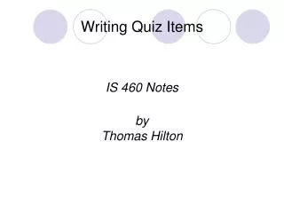
Colitis • Inflammation of the colon • Image – thickening of mucosal wall and loss of haustral markings • Diverticulum • outpouchingof the mucosal wall resulting from herniation of the inner wall of the colon. • Image – jagged or sawtooth appearance of the mucosa
Neoplasm • tumors in large intestine. • Image - narrowness or tapering of lumen “apple core” or “napkin-ring” lesions • Volvulus • twisting of a portion of the intestine on its own mesentery. • Image – corkscrew in appearance with air-filled distended region of the intestine
Intussusceptions • telescoping of one part of the bowel into another. • Image – mushroom-shaped dilation at the distal aspect of the intussusception with little or no gass passing beyond it.
Diverticulae Apple core Colitis Volvulus “coffe bean” Intussusceptions
Preparation of the Patient • The final objective is that the section of alimentary canal to be examined must be empty. 2 – classes of Cathartics (laxative) • Irritant cathartic – castor oil • Saline cathartic – magnesium citrate or sulfate
Contraindications to Cathartics • Gross bleeding • Severe diarrhea • Obstruction • Inflammatory lesions (appendicitis)
Contrast Media • High – density Barium Sulfate • It is excellent for use in double-contrast studies of the alimentary tract in which uniform coating of the lumen is required. • Air contrast • Carbon dioxide may also be used because it is more rapidly absorbed than nitrogen of air when evacuation.
Mixture of Barium suspensions • 12 % - 25% weight / volume – Single contrast • 75% - 95% weight / volume – Double contrast
Barium Containers • Closed system type enema • Open system type enema
Enema Tips • 3 – common enema tips • Plastic disposable • Rectal retention • Air contrast retention
Enema tips insertion • Sims position – relaxes the abdominal muscles and decreases pressure within the abdomen.
Summary of Enema tip insertion • Describe the tip insertion to pt. • Place pt in sims position. (pt should lie on the left side, with the right leg flexed at the knee and hip • Shake and inspect the enema container to provide good mixture. Allow the barium to flow through the tubing and from tip to remove any air in the system
Wearing gloves, coat enema tip with water-soluble lubricant.(KY jelly or any sterile lubricant) On expiration, direct enema tip toward the umbilicus proximally 1 to 1.5 inches After initial insertion, advance up superiorly and slightly anteriorly. Do not force enema tip.
Tape tubing in place to prevent slippage. Do not inflate unless directed by radiologist Ensure IV pole/enema bag is no more than 24 inches (60cm) above the table. Ensure tubing stopcock is in the closed position and no barium flows into the pt.
Procedures • 3 – Types of Examinations of Colon • Single – contrast Ba. Enema • Double – contrast Ba. Enema • Defecogram
Cont… • Single – contrast • utilizes only a positive contrast medium. • Double – contrast • Difference is that in an examination there is both air and barium.