Download
1 / 24
240 likes | 371 Vues
NSLS-II Environmental Sciences Breakout Session. 2. 8. 6. 5. 2. What are the key scientific drivers? What experiments will NSLS-II enable that are not presently possible? What technical capabilities will these require? (Beamlines, endstations, undulators…) Estimate of community size.

E N D
2 8 6 5 2
What are the key scientific drivers? What experiments will NSLS-II enable that are not presently possible? What technical capabilities will these require? (Beamlines, endstations, undulators…) Estimate of community size. What detector requirements does this field have? Do these require R+D? What software and computing infrastructure requirements are there? (Control, data acquisition, analysis) Any particular accelerator requirements? Any particular conventional facility requirements? Report Summarizing what was learned will be sent to DOE by COB August 5th. NSLS-II Workshop Deliverables
Synchrotron Hard X-Ray Microprobe in Environmental Sciences Why an X-ray microprobe:Distinct advantages over many analytical techniques by allowing analyses to be done in-situ and/or in-vivo, for example being the ability to determine chemical speciation of a wide variety of toxic elements in moist soils and biological specimens with little or no chemical pretreatment, low detection limits, and minimal beam interaction. Multiple Complimentary Techniques • µXRF and elemental mapping: Spot XRF analyses of trace element composition. • µXAFS: Spot XANES and EXAFS determinations of oxidation state and speciation. • µXRD: In-situ phase identification and correlation with elemental and speciation information. • Fluorescence Microtomography: Internal 2D and 3D elemental imaging.
X-ray Focusing and Imaging – Current State of the Art Modified from Jörg Maser, 2006
X-ray Focusing and Imaging – Current State of the Art State of the art in x-ray imaging and focusing (2D focus): • Refractive Optics: δ ~ 50 nm (E = 21 keV) (Schroer, APL, 2005) • Reflective Optics: δ ~ 40 nm (E ~ 15 keV) (Mimura, JJAP 2005) • Diffractive Optics: δ ~ 15 nm, (E = 0.8 keV) (Chao, Nature, 2005) What is the ultimate resolution limit for x-ray focusing? • Diffractive optics: ~ 1 nm (Kang, 2006); Å feasible? • Reflective Optics: ~ 16 nm (KB), 3 nm (Wolter) (non-ML) • Refractive Optics: ~ 2 nm (β = 0, Schroer, 2005)
Advantages of KB Microfocusing Combined capabilities for small spot size, achromatic focusing, large gain and long working distance. • Achromatic focusing - focus/beam position is retained during an energy scan • Large gain - gains of > 105 achievable, high elemental sensitivity • Long working distance - simplifies use of detectors, optical viewing systems, special sample chambers, etc. • Disadvantage - beam sizes ~ 0.1 micrometer currently unachievable for hard x-rays KB Microfocusing System Designed by P. Eng (U. Chicago)
An example is beamline 2ID-D at the APS Zone plate based HXR µprobe 100 nm-width tin oxide nanobelt
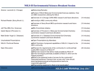
Synchrotron-based µ-XRF mapping, µ-XANES and µ-XRD of arsenic-rich gold mine tailings and lacustrine sediments from Yellowknife Bay, Canada Technique: X-ray Micro- Fluorescence, Spectroscopy, Diffraction Researchers: H. Jamieson, S. Walker, C. Andrade, (Queen’s University, Canada), A. Lanzirotti and S. Sutton (U. Chicago, CARS) Publication: Walker, S.R., Jamieson, H.E., Lanzirotti, A. and Andrade, C.F. (2005) Determining arsenic speciation in iron oxides: Application of synchrotron micro-XRD and micro-XANES at the grain scale. Canadian Mineralogist, v. 43, p. 1205-1224 Characterize As bearing solids in roaster residue and roaster-derived iron oxides in a subareal weathered tailings horizon Oxidation state and bonding mechanisms at the scale of individual particle (µ-XANES). Phase identification (µ-XRD) of individual grains. Objective is to distinguish hematite (a-Fe2O3) and maghemite (g-Fe2O3). Chemical mapping of individual grains (µ-XRF). Understand bioavailability, predict long-term stability, design remediation to ensure As immobility. hematite (a-Fe2O3) and maghemite (g-Fe2O3) Field of view 0.16mm x 0.10mm Complex zoning at micron scales
Synchrotron-based µ-XRF mapping, µ-XANES and µ-XRD of arsenic-rich gold mine tailings and lacustrine sediments from Yellowknife Bay, Canada Characterize As bearing solids in roaster residue and roaster-derived iron oxides in a subareal weathered tailings horizon Oxidation state and bonding mechanisms at the scale of individual particle (µ-XANES). Phase identification (µ-XRD) of individual grains. Objective is to distinguish hematite (a-Fe2O3) and maghemite (g-Fe2O3). Chemical mapping of individual grains (µ-XRF). Understand bioavailability, predict long-term stability, design remediation to ensure As immobility. H. Jamieson, S. Walker, C. Andrade, (Queen’s University, Canada), A. Lanzirotti and S. Sutton (U. Chicago, CARS)
Optical Image Pu L MicroXANES Pu “hot spots” located by microXRF mapping Influence of Plutonium Oxidation State on Long-Term Transport through a Subsurface Sediment • Pu(IV) and Pu(VI) were placed in Savannah River Site lysimeters (1980) exposed to natural weather conditions, with the intent of evaluating the long-term environmental fate of Pu. • Pu in the Pu(VI)-amended lysimeter traveled ~10 times faster (12.5 cm yr-1) than the Pu(IV)-amended lysimeter (1.1 cm yr-1) • MicroXANES showed Pu oxidation state was IV in IV-amended system (undetectable oxidized species) Yucca Mtn Tuff soak experiment M. C. Duff, D. Kaplan, Savannah River National Laboratory (SRNL), B. Powell, Clemson U., A. Lanzirotti and S. Sutton (U. Chicago, CARS)
Interplanetary Dust Particles 1 µm Potentially most primitive solar system solids Meteoritic material least altered by atmospheric entry Hosts of interstellar grains • Total mass ~ 30 picogram (trillionths of a gram)
10 mm IDPs: Fluorescence Microtomography Are the volatile element enrichments indigenous or stratospheric contamination? Fluorescence tomography images show that volatile elements (Zn and Br) are not strongly surface-correlated, suggesting that these elements are primarily indigenous rather than from atmospheric contamination Information on the host phases of trace elements (e.g., Zn, the first element lost during entry heating, is isolated in a few spots, probably ZnS identified by TEM in IDPs; Sr in carbonate) Sutton, S.R., et al. (2000) Lunar Planet. Sci. XXXI,1857.
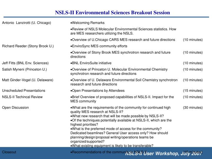
Ionomics: the study of how genes regulate ions in cells. Fe deficiency most common nutritional disorder in the world 2 billion (mainly in developing countries) are anemic A number of the key genes involved in iron uptake in plants have been identified. Armed with this knowledge, it should now be possible to engineer or breed plants with improved iron uptake abilities and in more bioavailable forms. Use synchrotron x-ray microprobe techniques to assign functions to metal homeostasis genes whose phenotypes could not be observed using volume-averaged metal analysis techniques Genomics to Ionomics: Metal homeostatis in plants Technique: X-ray Fluorescence Computed Micro-Tomography Researchers: T. Punshon and M. L. Guerinot (Dartmouth U.), A. Lanzirotti (U. Chicago, CARS) Publication: S. Kim, T. Punshon, A. Lanzirotti, L. Li, J. Alonso, J. Ecker, J. Kaplan and M. L. Guerinot (2006) Localization of Iron in Arabidopsis Seed Requires the Vacuolar Membrane Transporter VIT1, Science. Arabidopsis thaliana (Mouse-Eared Cress) genome sequenced (2000)
seed coat cotyledons radicle Absorption Tomograms
Col-O atvit1-1 Fe Mn Zn Mn Zn Fe Fluorescence Tomograms • Synchrotron x-ray fluorescence microtomography shows that the majority of iron is precisely localized to the provascular strands of the embryo. This localization of iron is completely abolished when the vacuolar iron uptake transporter VIT1 is disrupted, making vacuoles a promising target for increasing the iron content of seeds.