Download

1 / 33

E N D
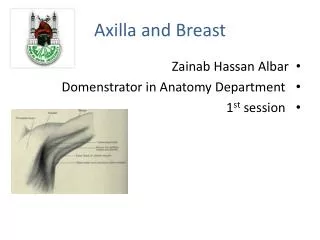
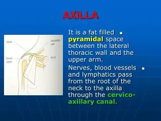
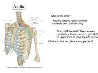
The axilla, or armpit, is a pyramidshaped space between the upper part of the arm and the side of the chest.Realize that the upper end, or apex, is directed into the root of the neck and is bounded anteriorly by the clavicle, posteriorly by the upper border of the scapula, and medially by the outer border of the first rib. The lower end, or base, is bounded anteriorly by the anterior axillary fold (formed by the lower border of the pectoralis major muscle), behind by the posterior axillary fold (formed by the tend of latissimus dorsi and the teres major muscle), and medially by the chest wall.
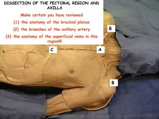
Carefylly remove the axillary fascia and fat and identify the axillary lymph nodes. Understand that the axillary lymph nodes are divided up into the following groups: (1)anterior or pectoral,(2)posterior orsubscapular, (3)central group, (4)lateral group, (5)apical group.Note the areas that they drain.
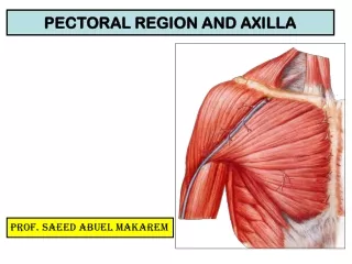
Walls of the Axilla. Identify the structures forming the walls of the axilla:
Anterior wall. This is formed by the pectoralis major, pectoralis minor, clavipectoral fascia, and subclavius muscle (the pectoral muscles and the clavipectoral fascia have already been removed).
Posterior wall.From above downward this wall is formed by the subscapularis, latissimus dorsi, and the teres major muscles.
Medial wall. This is formed by the upper four or five ribs and the intercostal spaces covered by the serratus anterior muscle.
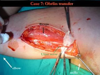
Lateral wall. This is formed by the coracobrachialis and biceps muscles in the bicipital groove of the humerus.
Axillary Sheath. The axillary artery (but not the vein) and the brachial plexus are enclosed in a fascial sheath derived from the prevertebral layer of deep cervical fascia in the neck. Identify the sheath. Clean the axillarty artery and its branches.
Axillary Artery. This commences at the outer border of the first rib as a continuation of the subclavian artery. Having passed through the axilla, it becomes the brachial artery at the lower border of the teres major muscle. It isarbitrarily divided into three parts by the pectoralis minor muscle that crosses it anteriorly.
Identify the following branches of the axillary artery: 1.Highest thoracic artery. This is a small artery that runs along the upper border of the pectoralis minor to the thoracic wall.
2.Thoracoacromial artery. This pierces the clavipectoral fascia and immediately divides into terminal branches. 3.Lateral thoracic artery. This runs along the lower border of the pectoralis minor to the thoracic wall.
4.Subscapular artery.This runs along the lower border of the subscapularis muscle. 5.Anterior and posterior circumflex humeral arteries.These wind around the anterior and posterior surface of the surgicalneck of the humerus, respectively.
Comfirm that the cephalic vein drains into the axillary vein. Now transect and remove the axillary vein and its tributaries.
Brachial Plexus. Identify and clean the branches of the brachial plexus. Note their close relationship to the axillary artery.
1.Identify the musculocutaneous nerve firstly as it pierces the coracobrachialis muscle. Note that this nerve supplies the muscle prior to piercing it.
2.Follow the musculocutaneous nerve proximally to the point where it arises from the lateral cord of the plexus. Identify the lateral root of the median nerve on the lateral side of the axillary artery.
3. Identify the lateral pectoral nerve arising from the lateral cord of the plexus.
4. Identify the medial root of the median nerve on the medial side of the axillary artery. Follow the median nerve down the lateral side of the axillary artery.
5. Follow the medial root of the median nerve proximally and identify the medial cord of the plexus. 6.Identify the remaining branches of the medial cord, namely, the medial pectoral nerve, the ulnar nerve, the medial cutaneous nerve of the forarm, and the medial cutaneous nerve of the arm.
7. The radial nerve is a large nerve lying posterior to the distal part of the axillary artery. Trace this nerve proximally and identify the axillary nerve, which is the other terminal branch of the posterior cord of the plexus.
8. The upper and lower subscapular nerves and the thoracodorsal nerve (middle subscapular nerve) arise from the posterior cord of the plexus.The upper subscapular nervesupplies the upper part of the subscapularis muscle, the lower subscapular nerve may be traced to the lower part of the subscapularis muscle and the teres major muscle, and the thoracodorsal nerve (middlesubscapular nerve)supplies the latissimus dorsi muscle.
Note that the medial cutaneous nerve of the arm usually communicates with the intercostobrachial nerve. This is the lateral cutaneous branch of the second intercostal nerve, which emerges from the second intercostal space. Identify the long thoracic nerve as it runs down the medial wall of the axilla and supplies the serratus anterior muscle. Having identified all the structures in the axilla,carefully clean them.
Study the origin of the serratus anterior muscle, which arises by eight digitations from the outer surface of the upper eight ribs. This is a large, flat muscle that runs posteriorly around the thoracic wall to beinserted into the anterior surface of the medial border of the scapula.
Clean the subscapularis muscle and note that it arises from the anterior surface of the scapula and is inserted into the lesser tuberosity if the humerus.
Follow the tendon of the latissimus dorsimuscle to its insertion into the floor of the bicipital groove of the humerus. Clean the teres major muscle and trace it to its insertion into the medial lip of the bicipital groove of the humerus.