Download
1 / 48
540 likes | 1.03k Vues
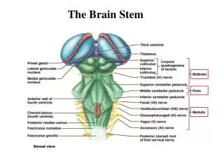
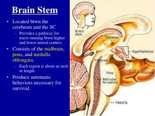
Brain stem. Pons – Midbrain. Pons. Located anterior to cerebellum 1 inch long Anterior surface is convex & shows transverse fibers that converge on each side to form middle cerebellar peduncle Located between the midbrain and medulla oblongata

E N D
Brain stem Pons – Midbrain
Pons • Located anterior to cerebellum • 1 inch long • Anterior surface is convex & shows transverse fibers that converge on each side to form middle cerebellar peduncle • Located between the midbrain and medulla oblongata • Contains the nuclei of cranial nerves V, VI, and VII
Gross appearance (anterior surface) • Basilar groove (midline)..lodges basilar artery • 5th nerve emerges from anterolateral surface (small motor (medial) and large sensory (lateral)) • 6th 7th &8th emerges at pontomedullary junction M→L
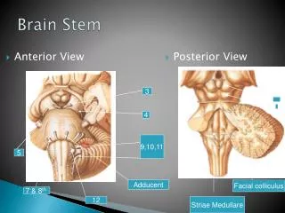
Pons….posterior view • Its hidden by from view by cerebellum • Forms the upper half of floor of 4th ventricle • Triangular in shape • Median sulcus • Medial eminence • Sulcus limitans • Facial colliculus (inf end of medial eminence)..Its produced by the root of facial nerve winding around the nucleus of abducent nerve
Internal structure of pons • Its divided by transversely running fibers of trapezoid body into: • Tegmentum (post part) • Basal part (ant part)
levels • Level through caudal part (facial colliculus) • Level through cranial part (trigeminal nuclei)
Level through caudal part (facial colliculus) • Facial nucleus • Abducent nucleus • MLF • Trapezoid body • Pontine nuclei and transverse fibers • Corticospinal and corticonuclear tracts • Medial leminiscus ,lateral leminiscus, spinal leminiscus • Spinal nucleus of trigeminal and its tract
Basilar part of pons contain small masses of nerve cells called pontine nuclei • Corticopontine fibers terminate in pontine nuclei • Axons of these cells give origin to transverse fibers of the pons which cross the midline and intersect the corticospinal & corticonuclear tracts, breaking them into small bundles • Transverse fibers enter MCP to cerebellum • This connection is the main pathway linking cerebellum to cerebral cortex
Level through cranial part (trigeminal nuclei) • Motor nucleus of trigeminal n • Main Sensory nucleus of trigeminal n (lateral) • SCP, MCP • Trapezoid body • Medial leminiscus ,lateral leminiscus, spinal leminiscus
The trapezoid body • is part of the acoustic pathway • Made up of fibers derived from cochlear nuclei
lateral lemniscus • a tract of axons in the brainstem that carries information about sound from the cochlear nucleus to the contralateral inferior colliculus of the midbrain • Cochlear nuclei----trapezoid body----lateral lemniscus----inf colliculus-----medial geniculate body-----auditory cortex
spinal leminiscus • anterior and lateral spinothalamic tracts • Spinotectal tracts
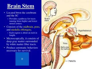
The Brain Stem – The Midbrain • Lies between the diencephalon and the pons • Central cavity – the cerebral aqueduct • Cerebral peduncles located on the ventral surface of the brain • Contain pyramidal (corticospinal) tracts • Superior cerebellar peduncles • Connect midbrain to the cerebellum
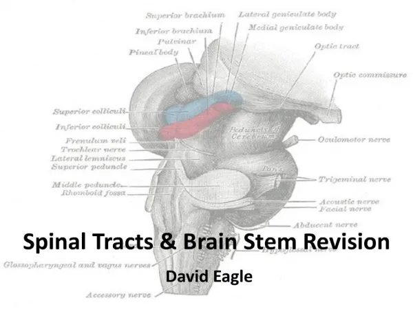
The Midbrain-posterior view • Corpora quadrigemina – the largest nuclei • Divided into the superior and inferior colliculi • Superior colliculi – nuclei that act in visual reflexes • Inferior colliculi – nuclei that act in auditory reflexes
Trochlear nerve emerges below the level of inf. Colliculus (from posterior surface) • Occulomotor nerve emerges at the level of sup. colliculus
Midbrain post view Sup. Collicullus Inf. Colliculus Sup.brachium (to lateral geniculate body) Inf. Brachium(to medial geniculate body) 4th emerges
Midbrain ant. View Interpeduncular fossa Crus cerebri 3rd nerve emerges from medial side of crus cerebri in the interpeduncular fossa
The Brain Stem – The Midbrain • Imbedded in the white matter of the midbrain • Two pigmented nuclei • Substantianigra – neuronal cell bodies contain melanin • Functionally linked to the basal nuclei • Red nucleus – lies deep to the substantianigra • Largest nucleus of the reticular formation
Cerebral peduncle is divided into crus cerebri (ant) & tegmentum (post) Tectum is post to cerebral aqueduct Substantianigra is situated between the tegmentum and cruscerebri
Level of inf. colliculus • Trochlear nucleus lies close to midline in the central gray matter • Trochlear nerves decussate in the superior medullary velum • Decussation of sup. cerebellar peduncles in the tegmentum • RF is lateral to decussation • Medial, spinal ,trigeminal & lateral leminisci • Substantianigra • Cruscerebri • RF • Mesencephalic nucleus of trigeminal (lateral to cerebral aqueduct) • MLF
Level at superior colliculus • Superior colliculus • Occulomotor nucleus • Occulomotor n emerges through red nucleus • MLF • Medial , trigeminal, spinal leminiscus (no lateral) • Red nucleus • Substantianigra • Cruscerebri • RF
Cruscerebri • Corticospinal & corticonuclear fibers (middle) • Frontopontine fibers (medial) • Temporopontine fibers (lateral) these descending tracts connect the cerebral cortex with spinal cord, cranial nerves nuclei, pons & cerebellum
Superior colliculus • Large nucleus of gray matter • Lies beneath corresponding surface elevation • Part of visual reflex • Connected to lateral geniculate body by sup. brachium
Inferior colliculus • Large nucleus of gray matter • Lies beneath corresponding surface elevation • Part of auditory pathway • Connected to medial geniculate body by inf. Brachium • Receives many terminal fibers from lateral leminiscus
Auditory pathway • Cochlear nuclei-----trapezoid body-----lateral leminiscus-----inferior colliculuus----- inferior brachium-----medial geniculate body
Lateral leminiscus • is a tract of axons in the brainstem that carries information about sound from the cochlear nucleus to various brainstem nuclei and ultimately the contralateral inferior colliculus of the midbrain
Substantianigra • Large motor nucleus • is a brain structure located in the midbrain • plays an important role in reward, addiction, and movement. • Substantianigra is Latin for "black substance" due to high levels of melanin • has connections with basal ganglia ,cerebral cortex, spinal cord • Concerned with muscle tone • Parkinson's disease is caused by the death of neurons in the substantianigra
Red nucleus • Rounded mass of gray matter • Situated bt cerebral aqueduct and substantia nigra • Reddish blue(vascularity & iron containing pigment) • Receive aff. Fibers from cerebral cortex,cerebellum,substantia nigra, thalamic nuclei, spinal cord • involved in motor coordination.