Download
1 / 48
580 likes | 1.19k Vues
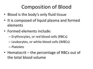
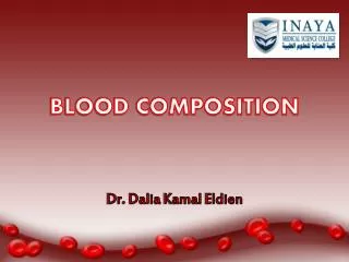
Blood Composition. Blood: a fluid connective tissue composed of Plasma Formed elements Erythrocytes (red blood cells, or RBCs) Leukocytes (white blood cells, or WBCs) Platelets. Blood Composition. Hematocrit Percent of blood volume that is RBCs 47% ± 5% for males

E N D
Blood Composition • Blood: a fluid connective tissue composed of • Plasma • Formed elements • Erythrocytes (red blood cells, or RBCs) • Leukocytes (white blood cells, or WBCs) • Platelets
Blood Composition • Hematocrit • Percent of blood volume that is RBCs • 47% ± 5% for males • 42% ± 5% for females
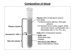
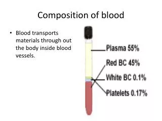
Formed elements Plasma • 55% of whole blood • Least dense component Buffy coat • Leukocytes and platelets • <1% of whole blood Erythrocytes 2 • 45% of whole blood • Most dense component 1 Centrifuge the blood sample. Withdraw blood and place in tube. Figure 17.1
Physical Characteristics and Volume • Sticky, opaque fluid • Color scarlet to dark red • pH 7.35–7.45 • 38C • ~8% of body weight • Average volume: 5–6 L for males, and 4–5 L for females
Functions of Blood • Distribution of ? • Protection against • Blood loss • Plasma proteins and clot formation • Infection • Antibodies • WBCs defend against foreign invaders
Blood Plasma • 90% water • Proteins are mostly produced by the liver • 60% albumin • 36% globulins • 4% fibrinogen
Platelets Erythrocytes Monocyte Neutrophils Lymphocyte Figure 17.2
2.5 µm Side view (cut) 7.5 µm Top view Figure 17.3
Erythrocytes • Structural characteristics contribute to gas transport • Shape—huge surface area • >97% hemoglobin • No mitochondria • No nucleus
bGlobin chains Heme group a Globin chains (a) Hemoglobin consists of globin (two alpha and two beta polypeptide chains) and four heme groups. (b) Iron-containing heme pigment. Figure 17.4
Hemoglobin (Hb) • O2 loading in the lungs • Produces oxyhemoglobin (ruby red) • O2 unloading in the tissues • Produces deoxyhemoglobin or reduced hemoglobin (dark red) • CO2 loading in the tissues • Produces carbaminohemoglobin (carries 20% of CO2 in the blood)
Hematopoiesis • Hematopoiesis; blood cell formation • Occurs in red bone marrow, girdle and proximal epiphyses of humerus and femur
Erythropoiesis:red blood cell production Stem cell Committed cell Developmental pathway Phase 1 Ribosome synthesis Phase 2 Hemoglobin accumulation Phase 3 Ejection of nucleus Reticulo- cyte Erythro- cyte Proerythro- blast Early erythroblast Late erythroblast Normoblast Hemocytoblast Figure 17.5
Regulation of Erythropoiesis • Too few RBCs leads to tissue hypoxia • Too many RBCs increases blood viscosity • Balance depends on • Hormonal controls • Adequate supplies of iron, amino acids, and B vitamins
Hormonal Control of Erythropoiesis • Erythropoietin (EPO) • Direct stimulus for erythropoiesis • Released by kidneys in response to hypoxia • Testosterone also enhances EPO production, resulting in higher RBC counts in males
Hormonal Control of Erythropoiesis • Causes of hypoxia • Hemorrhage or increased RBC destruction reduces RBC numbers • Insufficient hemoglobin (e.g., iron deficiency) • Reduced availability of O2 (e.g., high altitudes)
IMBALANCE Homeostasis: Normal blood oxygen levels 1 Stimulus: Hypoxia (low blood O2- carrying ability) due to • DecreasedRBC count • Decreased amountof hemoglobin • Decreasedavailability of O2 5 O2- carryingability of bloodincreases. IMBALANCE 4 Enhancederythropoiesisincreases RBCcount. 2 Kidney (and liver toa smaller extent)releaseserythropoietin. 3 Erythropoietinstimulates redbone marrow. Figure 17.6
Fate and Destruction of Erythrocytes • Life span: 100–120 days • Macrophages engulf dying RBCs in spleen • Iron is salvaged for reuse • Heme is degraded to yellow bilirubin • Liver secretes bilirubin (in bile)) into the intestines • Degraded pigment leaves the body in feces as stercobilin
1 Low O2levels in blood stimulate kidneys to produce erythropoietin. 2 Erythropoietin levels rise in blood. 3 Erythropoietin and necessary raw materials in blood promote erythropoiesis in red bone marrow. 4 New erythrocytes enter bloodstream; function about 120 days. 5 Aged and damaged red blood cells are engulfed by macrophages of liver, spleen, and bone marrow; the hemoglobin is broken down. Hemoglobin Heme Globin Bilirubin Amino acids Iron stored as ferritin, hemosiderin Iron is bound to transferrin and released to blood from liver as needed for erythropoiesis. Bilirubin is picked up from blood by liver, secreted into intestine in bile, metabolized to stercobilin by bacteria, and excreted in feces. Circulation Food nutrients, including amino acids, Fe, B12, and folic acid, are absorbed from intestine and enter blood. 6 Raw materials are made available in blood for erythrocyte synthesis. Figure 17.7
Erythrocyte Disorders • Anemia: abnormally low O2-carrying capacity • A sign rather than a disease itself • Blood O2cannot support normal metabolism • Accompanied by fatigue, paleness, shortness of breath, and chills
Causes of Anemia • Insufficient erythrocytes • Hemorrhagic anemia: acute or chronic loss of blood • Hemolytic anemia: RBCs rupture prematurely • Aplastic anemia: destruction or inhibition of red bone marrow
Causes of Anemia • Low hemoglobin content • Iron-deficiency anemia • Secondary result of hemorrhagic anemia or • Inadequate intake of iron-containing foods or • Impaired iron absorption
Causes of Anemia • Pernicious anemia • Deficiency of vitamin B12 • Lack of intrinsic factor needed for absorption of B12 • Treated by intramuscular injection of B12
Causes of Anemia • Sickle-cell anemia • Defective gene codes for abnormal hemoglobin (HbS) • Causes RBCs to become sickle shaped in low-oxygen situations
(a) Normal erythrocyte has normal hemoglobin amino acid sequence in the beta chain. 1 2 3 4 5 6 7 146 (b) Sickled erythrocyte results from a single amino acid change in the beta chain of hemoglobin. 1 2 3 4 5 6 7 146 Figure 17.8
Erythrocyte Disorders • Polycythemia: excess of RBCs that increase blood viscosity • Results from: • Polycythemia—bone marrow cancer • Secondary polycythemia—when less O2 is available (high altitude) or when EPO production increases • Blood doping
Differential WBC count (All total 4800– 10,800/l) Formed elements Platelets Granulocytes Neutrophils (50–70%) Leukocytes Eosinophils (2–4%) Basophils (0.5–1%) Erythrocytes Agranulocytes Lymphocytes (25 – 45%) Monocytes (3 – 8%) Figure 17.9
Leukopoiesis • Production of WBCs • Stimulated by chemical messengers from bone marrow and mature WBCs
Stem cells Hemocytoblast Lymphoid stem cell Myeloid stem cell Committed cells Myeloblast Myeloblast Myeloblast Monoblast Lymphoblast Developmental pathway Promonocyte Promyelocyte Promyelocyte Promyelocyte Prolymphocyte Eosinophilic myelocyte Basophilic myelocyte Neutrophilic myelocyte Basophilic band cells Eosinophilic band cells Neutrophilic band cells Monocytes Neutrophils Eosinophils Basophils Lymphocytes (a) (e) (b) (c) (d) Agranular leukocytes Some become Granular leukocytes Some become Figure 17.11
Leukocyte Disorders • Leukopenia • Abnormally low WBC count—drug induced • Leukemias • Cancerous conditions involving WBCs • Named according to the abnormal WBC clone involved • Myelocytic leukemia involves myeloblasts • Lymphocytic leukemia involves lymphocytes • Acute leukemia involves blast-type cells and primarily affects children • Chronic leukemia is more prevalent in older people
Leukemia • Bone marrow totally occupied with cancerous leukocytes • Immature nonfunctional WBCs in the bloodstream • Death caused by internal hemorrhage and overwhelming infections • Treatments include irradiation, antileukemic drugs, and stem cell transplants
Platelets • Form a temporary platelet plug that helps seal breaks in blood vessels • Circulating platelets are kept inactive and mobile by NO and prostacyclin from endothelial cells of blood vessels
Stem cell Developmental pathway Hemocyto- blast Promegakaryocyte Megakaryoblast Megakaryocyte Platelets Figure 17.12
Hemostasis • Fast series of reactions for stoppage of bleeding • Vascular spasm • Platelet plug formation • Coagulation (blood clotting)
Vascular Spasm • Vasoconstriction of damaged blood vessel • Triggers • Direct injury • Chemicals released by endothelial cells and platelets • Pain reflexes
1 Step Vascular spasm •Smooth muscle contracts, causing vasoconstriction. 2 Step Platelet plug formation •Injury to lining of vessel exposes collagen fibers; platelets adhere. Collagen fibers •Platelets release chemicals that make nearby platelets sticky; platelet plug forms. Platelets 3 Step Coagulation •Fibrin forms a mesh that traps red blood cells and platelets, forming the clot. Fibrin Figure 17.13
Phase 1 Intrinsic pathway Extrinsic pathway Tissue cell trauma exposes blood to Vessel endothelium ruptures, exposing underlying tissues (e.g., collagen) Platelets cling and their surfaces provide sites for mobilization of factors Tissue factor (TF) XII Ca2+ XIIa VII XI XIa VIIa Ca2+ IX IXa PF3 released by aggregated platelets VIII VIIIa TF/VIIacomplex IXa/VIIIacomplex X Xa Ca2+ V PF3 Va Prothrombin activator Figure 17.14 (1 of 2)
Prothrombin activator Phase 2 Prothrombin (II) Thrombin (IIa) Phase 3 Fibrinogen (I) (soluble) Ca2+ Fibrin (insoluble polymer) XIII XIIIa Cross-linked fibrin mesh Figure 17.14 (2 of 2)
Thromboembolytic Conditions • Prevented by • Aspirin • Antiprostaglandin that inhibits thromboxane A2 • Heparin • Anticoagulant used clinically for pre- and postoperative cardiac care • Warfarin • Used for those prone to atrial fibrillation
Bleeding Disorders • Hemophilias include several similar hereditary bleeding disorders • Hemophilia A: most common type (77% of all cases); due to a deficiency of factor VIII • Hemophilia B: deficiency of factor IX • Hemophilia C: mild type; deficiency of factor XI • Symptoms include prolonged bleeding, especially into joint cavities • Treated with plasma transfusions and injection of missing factors
ABO Blood Groups • Types A, B, AB, and O • Based on the presence or absence of two agglutinogens (A and B) on surface of RBCs • Blood may contain anti-A or anti-B antibodies (agglutinins) that act against transfused RBCs with ABO antigens not normally present
Rh Blood Groups • Anti-Rh antibodies are not spontaneously formed in Rh– individuals • Anti-Rh antibodies form if an Rh– individual receives Rh+ blood • A second exposure to Rh+ blood will result in a typical transfusion reaction • RhoGAM containing anti-Rh can prevent the Rh– mother from becoming sensitized
Serum Blood being tested Anti-A Anti-B Type AB (contains agglutinogens A and B; agglutinates with both sera) RBCs Type A (contains agglutinogen A; agglutinates with anti-A) Type B (contains agglutinogen B; agglutinates with anti-B) Type O(contains no agglutinogens; does not agglutinate with either serum) Figure 17.16