Download
1 / 44
470 likes | 597 Vues
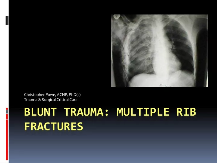
Christopher Powe, ACNP, PhD(c) Trauma & Surgical Critical Care. Blunt Trauma: Multiple Rib Fractures. OBJECTIVES. The participants will be able to: Articulate the epidemiology of ribs fractures associated with blunt trauma

E N D
Christopher Powe, ACNP, PhD(c) Trauma & Surgical Critical Care Blunt Trauma: Multiple Rib Fractures
OBJECTIVES The participants will be able to: • Articulate the epidemiology of ribs fractures associated with blunt trauma • Articulate the pathophysiology of multiple rib fractures associated with blunt trauma • Describe the management of multiple rib fractures associated with blunt trauma
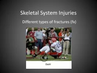
Introduction • Multiple Rib Fractures • Most common injury in blunt chest trauma • 10% patients have one or more rib fractures • Rarely life-threatening • CAN be a sign of more severe visceral injuries • Mechanism • Most common in elderly • Fall from height or standing position • Adults • Most common Motor Vehicle Crashes (MVC) • Youth • Recreational or athletic activities
Pathophysiology • Chest Wall • Protects underlying sensitive structures / organs • Provides hard osseous protection (i.e. ribs, clavicles, sternum and scapula • Intact chest wall necessary for normal respiration • Compromised Respiration by multiple mechanisms • Pain results in splinting, atelectasis and pneumonia • Flail chest interferes with costovertebral and diaphragmatic muscle excursion respiratory insufficiency
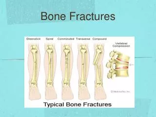
Pathophysiology • Penetration • Fractured ribs may cause HTX or PTX • Commonly fx at point of impact or posterior angle • (structurally at their weakest point) • Ribs 4-9th are most commonly injured • Vulnerability • Thinnest / weakest portion of 1st rib groove of subclavian artery • 1st rib injury in MVC is violent contraction of scalene muscles by sudden forward movement of head / neck
Epidemiology Frequency • Dramatically underreported • >2 million blunt mechanisms of injury occur annually as MVC • Chest injury 67-70% of those
Epidemiology • International • Prevalence is linked to underlying cause of trauma • More common in countries with higher incidence of MVC • Mortality/Morbidity • Rib f(x) not usually dangerous alone • Pneumonia / atelectasis largest risk • Morbidity correlates with degree of injury to underlying structures
Epidemiology • Mortality / Morbidity • Mortality rates of 12% in one study of rib f(x) • 94% had associated injuries / 32% had a HTX/PTX • Ziegler, W. J Trauma, Dec 2004. • > 50% required operative or ICU care • Average blood loss was 150-200ml per rib f(x)
Epidemiology • Retroprospective Study • 99 elderly patients • 16% adverse events • 2 deaths • Adverse events ( ARDS, pneumonia, intubation, transfer to ICU for hypoxemia or death • Lotifpour, S. West J Emerg Med May 2009 • Risk Factors • Age > 85 • Initial SBP < 90 • HTX / PTX • 3 or more unilateral rib fractures • Pulmonary contusions • Risk factors 100% sensitivity for predictability
Epidemiology • Elderly • Most common injury in blunt chest trauma • Each additional rib fracture increases mortality by 19% • Increases incidence of pneumonia by 27% • First Rib F(x) • Rarest of all • Once thought harbinger of severe trauma • Suspected of much higher impact force • These findings are now under suspicion • No real data to support
Epidemiology • First Rib F(x) • For now.. Should raise high suspicion for severe injury • Mortality rates >36% reported w/associated 1st rib fracture • Associated Injuries with 1st Rib F(x) • Lung parenchyma • Ascending aorta • Subclavian artery • Brahcial plexus • Delayed vessel thrombosis • Tracheobronchial fistula • Thoracic outlet syndrome
Epidemiology • Race • No supporting data • With exception of general trends associated with types of trauma • Sex • No supporting data • With exception of stratification of certain types of trauma and risk-taking behaviors
Epidemiology • Age • Children • Less likely to sustain rib fractures in chest trauma • More elastic ribs • More likely with trauma to chest/abd without rib f(x) • Rib f(x) in children is MORE OMINOUS SIGN of serious injury • Bruising near a f(x) site is uncommon in children (9.1%) • Child Abuse • Consider in absence of severe mechanism of injury • Multiple stages of healing • Rib f(x) in children <2 y/o have 83% incidence of abuse
Epidemiology • Age • Elderly more prone than young • Pulmonary sequelae including atelectasis, pneumonia and respiratory arrest more likely in elderly • Cardiopulmonary disease increases morbity/mortality in patients >65 y/o.
History • Description of pre-hospital scene by paramedics • MVC • Deformation of steering wheel • Activation of seatbelts/airbags • Associated with multiple rib fracutreus • Coughing • Rib fractures HAVE been reported with coughing w/out significant trauma • Athletes • high force, recurrent movements at risk for stress fractures of ribs
Physical Assessment • Tenderness to palpation • Chest wall deformity • Paradoxical chest wall excursion • FLAIL CHEST • Large segment of ribs not attached to spine • Ribs f(x) in at least 2 places on each rib • F(x) sites move in response to intrathoracic pressure changes NOT to intercostal contractions
Physical • Respiratory Insufficiency • Cyanosis • Tachypnea • Retractions • Uses of accessory muscles • F(x) of lower ribs • Assessment for abdominal tenderness • Be highly suspicious for intra-abdominal injury • Solid – and Hollow- organ injury
Diagnosis • Plain film imaging • May or may not provide useful imaging • “Clinical Diagnosis” may be utilized if mechanism and clinical presentation match • Major blunt trauma will require CT Imaging • Radiographs depict: • Bony trauma • Rib f(x) • Pleural space • Lungs • Extrapleural space • Mediastinum,heart • Great vessels and spine
Imaging • Anterior Plain Film • Ribs 1-12 • Note 12th Rib does not articulate with anteriorly • Ribs articulate anteriorly with costochondral junction
Imaging • Posterior Views • Posterior ribs are commonly injured along with scapula in MVC and/or ejection
Imaging • Fractures of 10-12th ribs • Hemorrhage of adrenal glands associated risk of these fractured ribs • Also associated with renal and splenic injury • Associated with vertebral lumbar and thoracic spine Presence of right PTX (blue arrow) with tracheal shift. (Black arrow) demonstrate posterior rib fracture.
Imaging • CT Chest • Demonstrates multiple left-sided rib fractures with LARGE PTX
Imaging • Posterior Rib F(x) • Left PTX (white arrows) with displaced posterior rib fracture • Note posterior left pulmonary contusion and atelectasis
Imaging • Flail Chest • MVC in ED • Left lateral chest rib fractures a (black arrows) • Metal artifacts have obscured additional rib fractures (blue arrows)
Imaging • Post/Lat Rib Fractures • Multiple posteriorlateral rib f(x) are noted
Imaging • Fall • Elderly female with severe left lateral chest wall pain • s/p fall • Left lateral rib fracture (arrow) that may be seen on a standard AP chest radiograph
Imaging • Left Rib Fractures • Left lateral rib fractures (white arrows) • Left lateral subcutaneous gas pattern dissecting along the left chest wall • High suspicion for PTX or parynchemal injury
Imaging • Rib F(x) with PTX • Severe blunt chest wall trauma • Left chest wall air (yellow arrow) • Small left PTX (blue arrow) • Left pulmonary contusion
Imaging • Left Rib F(x) • Opacity left lateral upper lobe (arrows) • Consistent with pulmonary contusion • Left lateral rib fractures
Imaging • 3D Imaging of Posterior Rib Fx • Utilized for more thorough evaluation and surgical options
Imaging • 3D Imaging left Rib F(x)
Imaging • This type of imaging also assists surgeons in determination of surgical options
Management • Initial Goals • Primary goal is pain relief • Adequate clearing of secretions • Isolated rib f(x) may be managed on an outpatient basis with oral analgesics • Outpatient Management • Topical Lidocaine – Lidoderm patchs • Incentive spirometer • Use of splinting techniques • NSAIDS – Ketoralac • Analgesics - Opiates
Management • Inpatient • Patient-controlled anesthesia may allows pain relief withou inhibition of respiratory drive • Intercostal nerve blocks without respiratory depression • Rib belts or binders: • May control pain • Also associated with atelectasis, hypoventilation and pneumonia • We avoid this technique at all costs
Management - Medication • Pain control is Mainstay • Meta-analysis of 8 studies (232 patients) DID NOT demonstrates significant benefit of epidural analgesia on mortality, ICU or hospital LOS compared with other analgesic modalities. • Mechanical Ventilation • Thoracic epidural analgesia with local anesthetics did demonstrate shorter intubation periods • Hypotension was reported as significant finding associated with epidural analgesia
Management - Medication • Accepted Mainstays of Treatment • Non-steroidal Anti-Inflammatory • Oral Narcotic Agents
Management - NSAIDS • These agents utilized for mild to moderate pain • Ibuprofen – first –line drug of choice • Ketoprofen – administer small doses initially to patietns with lower body weight • Naproxen – inhibits inflammatory reactions and pain by decreasing enzyme cyclooxygenase resulting in decreased prostaglandin synthesis
Management - Analgesics • Pain control is essential and use of analgesics in the acute setting is an appropriate and accepted standard of care. • Acetaminophen – alternative for NSAID hypersensitivity • Hydrocodone and acetaminophen • Oxycodone and acetaminophen • Hyodrocodone and ibuprofen • Morphine Sulfate –may provide both anxiolytic and analgesic affects and may be titrated.
What’s New • Early Rib-Plating vs Late Rib-Plating • In the past, patients with traumatic rib rib fractures >3 months old were considered for plating • Criteria based on deformity, non-union of the fracture and chronic pain. • Today • Ongoing studies currently being conducted • Question: Efficacy of early rib plating in the acute setting
What’s New • Current Academic Discussions • Who is a candidate for acute rib-plating? • Risks of operating on a polytrauma patient for acute rib plating? • Who should perform acute rib plating?
What’s New • Minimally Invasive Surgical Options • Currently there is much debate among researchers on who is a candidate for surgical rib plating • They all agree there is a small sub-set of patients that my benefit from this procedure • Certainly, the medical device companies believe ALL (325,000 patient annually) are candidates! • Agreed upon criteria for candidacy: • 3 or more rib fractures • Flail segments • Gross deformity • Chronic pain
CONCLUSION & OBJECTIVES The participants will be able to: • Articulate the epidemiology of ribs fractures associated with blunt trauma • Articulate the pathophysiology of multiple rib fractures associated with blunt trauma • Describe the management of multiple rib fractures associated with blunt trauma