Download
1 / 36
360 likes | 513 Vues
Chapter 20 – The Lymphoid System and Immunity . Functions of lymphoid system . Production (along with red bone marrow) and distribution of lymphocytes Fluid balance Cardiovascular system delivers more fluid to peripheral tissues (via capillaries) than it drains away

E N D
Functions of lymphoid system • Production (along with red bone marrow) and distribution of lymphocytes • Fluid balance • Cardiovascular system delivers more fluid to peripheral tissues (via capillaries) than it drains away • Excess fluid enters lymphatic system, and is then returned to cardiovascular system
Components of lymphoid system • Lymph • Has lower concentration of proteins than plasma • Lymphatic vessels • Collect fluid from tissues and return to venous system • Lymphoid tissues and organs
Lymphatic capillaries • Smallest vessel type • Originate as pockets (not continuous as in cardiovascular system) • Endothelial cells overlap and form valves • Allows entry into lymphatic system, but prevents re-entry into intercellular spaces • Lacteals are large capillaries in small intestine • Involved with lipid transport
Small lymphatic vessels • Lead toward body’s trunk • Similar in structure to veins • Have valves, which form bulges in the vessel • Valves are closer together than in veins
Major vessels • Superficial lymphatics • Dermal layer of skin; areolar layer of GI tract; respiratory, urinary, and reproductive systems • Deep lymphatics • Accompany arteries and veins of skeletal muscles, deeper organs • Superficial and deep converge to form lymphatic trunks
Major vessels cont • Trunks converge to form: • Thoracic duct • Collects lymph from body inferior to diaphragm and left side of body superior to diaphragm • Cisterna chyli – sac-like area at base of thoracic duct • Receives lymph from lower regions • Right lymphatic duct • Collects lymph from right side of body superior to diaphragm
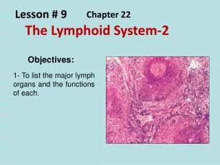
Lymphoid organs and tissue • Organs • Lymph nodes • Thymus • Spleen • Tissues • MALT (mucosa-associated lymphoid tissue) • Tonsils
Lymph nodes • Kidney bean shape • Afferent lymphatic carries lymph into nodes; efferent lymphatic carries out • Purifies lymph before returning to venous circulation • 99% of foreign antigens are removed • Macrophages in walls remove debris • Serve as antigen-presenting cells • Largest nodes are cervical, axillary, and inguinal
Thymus • Largest (relative to body size) during young childhood (1 – 2 yrs) • After puberty, starts to become fibrous • Involution • Two lobes • Septa divide each into smaller, multiple lobules • Outer region = cortex; inner region = medulla • Site of T cell maturation
Spleen • Removes abnormal blood cells from phagocytosis • Stores iron (recycled from blood cells) • Initiation of immune responses by T and B cells to antigens in circulating blood • Reticular connective tissue • Red pulp – contains RBCs • All circulating components of blood, and macrophages • White pulp – contains lymphocytes (stains purple)
Lymphoid tissues • MALT (mucosa-associated lymphoid tissue) • Peyers patches in intestinal walls; walls in vermiform appendix • Tonsils • Pharyngeal – singular (“adenoid”) • Palatine – paired • Lingual – paired
Innate defenses • First line of defense against infection • Bacteria, viruses, other pathogens • Present before an exposure occurs • Non-specific
Innate defenses cont • Skin • Lysozyme – digests bacterial cell walls • Mucous membranes • Traps microbes; stomach acids kill most bacteria • Neutrophils/macrophages • Engulf microbes by phagocytosis • Natural killer cells • Attack cancer cell and viral-infected cells by releasing chemicals to promote apoptosis
Innate defenses cont • Interferons • Produced by viral-infects cells that help other cells resist infection • Complement • ~30 proteins in plasma • Circulate in an inactive form – activated by exposure to microbes • Leads to lysis of invader and an inflammatory response
Innate defenses cont • Inflammatory response • Histamine released by damages tissue causes nearby blood vessels to dilate and become ‘leaky’ • Complement attracts phagocytes to area • Clotting proteins and platelets form local clot to seal off infected area
Acquired immunity • Fully develops AFTER exposure to a pathogen • Can amplify non-specific responses (inflammation, complement) • Responds to the presence of foreign antigens • Increase the number of cells that fight invader, and produce defense proteins called antibodies • Has memory • A second exposure to a previously encountered antigen is faster and stronger • Acquired by: • Natural exposure • Vaccination/immunization
Types of acquired immunity • Active • Person’s own immune system actively produces antibodies • Either due to natural exposure or artificial (vaccine) • Passive • Receive pre-made antibodies • Maternal antibodies to fetus through placenta • Breast milk to babies • Shot of antibodies after high-risk exposure • Lasts only as long as antibody “lifespan” • Weeks to months
Lymphocytes – B cells • B cells differentiate in bone marrow • Humoral immunity • Secrete antibodies which circulate in blood and lymph • Defend against bacteria and viruses in body fluids • Antibodies join with antigen • Complex recognized by macrophages
Antigens • Have specific regions where antibodies bind to them • Antigenic determinant or epitope • Small region on antigen’s surface • Recognized by antibody • Single antigen may be recognized by different antibodies at different sites
Antibody structure • 4 polypeptide chains • Each arm of Y has 2 variable regions that form a specific antigen recognition site • C region sequences determine class of antibody
Classes of antibodies • IgG • Most abundant • Only one that can cross the placenta • Fights bacteria, viruses, toxins; activates complement • IgM • First type produced by newborn • Antibodies for ABO antigens; activates complement • IgA • Found in saliva, tears, colostrum, mucous membranes • Primary defense against local infections of mucous membranes • Prevents attachment of viruses and bacteria to epithelial cells
Classes of antibodies cont • IgD • Cell membranes of B lymphocytes • Antigen receptor for initiation of B cell differentiation • IgE • Involved in inflammation, allergic responses, parasitic infections • Binds to mast cells and basophils • Triggers release of histamine and other inflammation & allergy mediators
Lymphocytes – T cells • Differentiate in thymus • Cell-mediated immunity • Circulate in blood and attack cells that are infected • Also protects against fungal and protist invaders • Aid in elimination of cancerous cells
T cells • Helper T cells (CD4) • Interact with other WBCs that function as antigen-presenting cells • Activation of helper T stimulates other cells of immunity • Cytotoxic T cells (CD8) • Attack body cells that are infected by pathogens
Hypersensitivity • Excessive or inappropriate activation of the immune response • The body is damaged by the immune response, rather than by the antigen • Type I hypersensitivity • Commonly called ‘allergic reactions’ • Mediated by IgE
Anaphylaxis • Systemic response to the inflammatory mediators released in type I hypersensitivity • Histamine, acetylcholine, kinins, leukotrienes, and prostaglandins all cause vasodilation • Acteylcholine, kinins, leukotrienes, and prostaglandins all can cause bronchoconstriction
Autoimmune diseases • Immune system attacks self-antigens • Normally, self-reactive immune cells are killed in the lymphoid organs or suppressed by regulatory T cells • In autoimmunity, this self-tolerance breaks down • Immune system destroys body tissues • Anti-tissue antibodies appear in blood (ex: anti-thyroid antibodies)
HIV – human immunodeficiency virus • HIV is a retrovirus • Has RNA instead of DNA as its genetic material • Uses the enzyme reverse transcriptase to make DNA from RNA • Binds to CD4 receptors
HIV cont • New viral particles can be shipped from cell via exocytosis • Mutation then causes HIV to destroy helper T cells (cells lyse) • AIDS – helper T cell population is dangerously low • Affects humoral and cell mediated immunity