Download
1 / 16
320 likes | 541 Vues
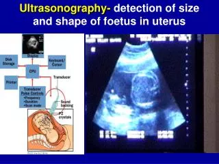
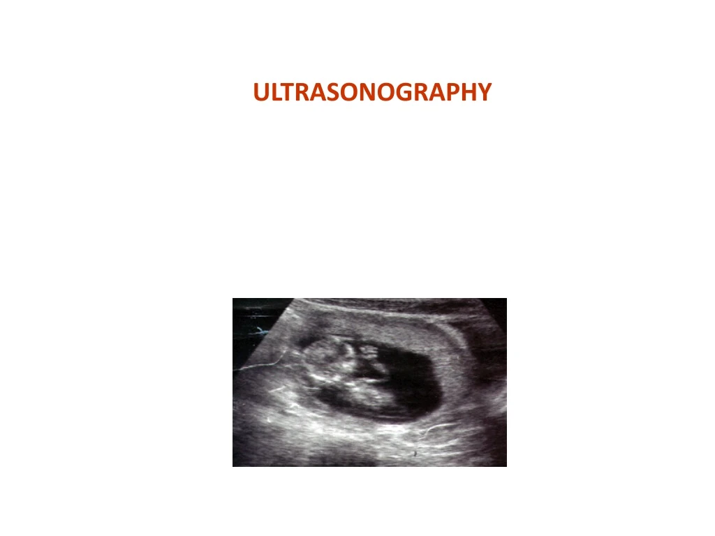
ULTRASONOGRAPHY. What is Ultrasound?. High frequency, inaudible sound waves of 1-10 MHz Audible frequency is 20-20,000 Hz Reflection of ultrasound occurs between substances of different acoustic impedance Product of velocity of sound and density of substance

E N D
What is Ultrasound? • High frequency, inaudible sound waves of 1-10 MHz • Audible frequency is 20-20,000 Hz • Reflection of ultrasound occurs between substances of different acoustic impedance • Product of velocity of sound and density of substance • Ultrasound waves unable to pass through vacuum and transmission in air is poor
Ultrasound • Ultrasound uses high frequency sound waves and their echoes. • The ultrasound machine transmits high-frequency sound pulses. • The sound waves hit a boundary between tissues (e.g. between fluid and soft tissue, soft tissue and bone). • Some of the sound waves get reflected back to the probe, while some travel on further until they reach another boundary and get reflected.
Principle of Ultrasound Electricity Transducer U/S waves Scan converter Transducer Tissue Cathode tube Image of Object
Ultrasound • Transducer sends and receives high frequency sound waves • Transducer must be in contact with structure being examined • Ultrasound gel or obstetric lube used to make contact complete.
How ultrasounds generated and detected? • High voltage to piezoelectric crystals generates waves of specific frequency • Higher the frequency, better the details (better differentiation), but poor penetration (depth) • Lower the frequency, poorer the details, better penetration
Wave length • Higher frequency = shorter wavelength • Shorter wavelength = better resolution • Better resolution = view smaller objects with more detail • Shorter wavelength = less penetration • Less penetration = view objects close to surface • Rectal exams 5 MHz transducer
Wave length • Lower frequency = Longer wavelength • Longer wavelength = less resolution • Less resolution = less detail • Longer wavelength = more penetration • More penetration = view deeper objects • Transabdominal 3 MHz transducer
7 mHz 5mHz 3mHz
Transducers • Crystals vibrate at high frequency producing sound waves • The sound waves are transmitted and received by the transducer • The transducer is identified by the crystal array. • The array is the way in which the crystals are arranged • Linear array • Convex array
Transducers • Piezoelectric material, Lead Zirconate Titanium • Frequency of 1 to 10 MHz is used • Linear transducers • 64-256 no of crystals • Sealed to avoid moisture
How is interpretation done? • Anechoic Black (Fluids) • Hypoechoic Grey (Soft Tissue) • Hyperechoic White (Bones)
Ultrasound Wave Transmission Transducer Sends and receives ultrasound waves WATER SOFT TISSUE BONE /AIR/ METAL