Download
1 / 103
1.19k likes | 1.77k Vues
Chapter 28 DNA Metabolism: Replication, Recombination, and Repair. Outline. The DNA replication process. Properties of DNA polymerases. Why are there several DNA polymerases ? Eucaryotic DNA replication. Replication of the ends of chromosomes. Replication of RNA genomes.

E N D
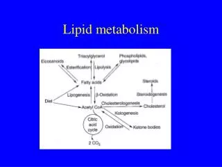
Chapter 28DNA Metabolism: Replication, Recombination, and Repair
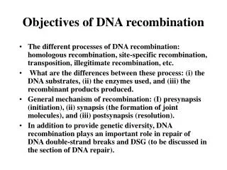
Outline • The DNA replication process. • Properties of DNA polymerases. • Why are there several DNA polymerases ? • Eucaryotic DNA replication. • Replication of the ends of chromosomes. • Replication of RNA genomes. • Genetic recombination: shuffling genetic information. • DNA repair. • The molecular basis of mutation. • Proteins as genetic agents.
The Dawn of Molecular Biology April 25, 1953 • Watson and Crick: "It has not escaped our notice that the specific (base) pairing we have postulated immediately suggests a possible copying mechanism for the genetic material." • The mechanism: Strand separation, followed by copying of each strand. • Each separated strand acts as a template for the synthesis of a new complementary strand.
28.1 DNA Replication • DNA replication is semiconservative – one of the two original strands is conserved in each progeny molecule. Replication is neither conservative nor dispersive. • DNA replication is bidirectional – it proceeds in both directions from the starting point using two replication forks. • Replication requires unwinding of the DNA helix (using a helicase) and relieving stress due to unwinding (using a topoisomerase).
Features of DNA Replication • DNA replication is semidiscontinuous. • The leading strand copies continuously. • The lagging strand copies in segments (discontinuous). The lagging strand is formed from Okazaki fragments, which are joined together to form a continuous strand. • Synthesis of both new strands occurs in the 5' 3' direction using a 3' 5' template
28.1 DNA Replication Figure 28.1 DNA replication Three Steps: 1. Initiation: Ori site in E.coli = OriC. This is a 245 bp highly conserved sequence. 2. Polymerization: Chain elongation in the 5'-3' direction. 3. Termination: The “ter region” is a ~ 350 kbp seq. that is 180o from OriC.
E. Coli OriC site OriC in E.coli is where replication is initiated. There are three-13 bp AT rich concensus sequences on 5' end of OriC, (5'-GATCTNTTNTTTT). These AT rich sites have only two H-bonds per bp (weak binding) and separate to form a bubble. AT rich DnaA binding 5' =13=13=13======9====9====9====9====9== 3' To the right of the AT rich region are five-9 bp sites that have opposing sequences (5'-TTATCCACA) and bind as many as 10-20 DnaA proteins (the initiation factor) to form a nucleosome-like structure. These five sites bind DnaA and require hydrolysis of ATP to open the replication bubble in the AT rich region.
E. Coli DNA Priming Events HU a histone-like protein prevents DnaA from binding at sites other than OriC. When the loop is open DnaB, a helicase, binds at each fork as a complex (DnaB6•DnaC6•ATP6). DnaC is a chaperone for DnaB and DnaT assists forming the pre-priming complex. DnaC leaves when ATP hydrolyzes. DnaB continues to unwind increasing the bubble size displacing DnaA as it moves. SSB binds to ssDNA in the open bubble to prevent annealing. Topo II binds ahead of the fork to relieve stress.
E. Coli DNA Priming Events PriA, PriB and PriC enter the bubble along with DnaG (DNA primase). This completes the primosome which makes RNA primers. Topo II and SSB are not part of the primosome. DNA primase does not need a primer to begin synthesis of RNA primers. The primers (10-30 bp) begin at the center base of any GTT sequence. Only one primer is needed on the leading strand. After this is made, the primosome moves to the lagging strandand a new primer is started about every 1000 bp.
Figure 28.2 Bidirectional Replication Comparison of labeling during unidirectional versus bidirectional replication. An autoradiogram of E. coli chromosome replication.
E. Coli Features of Replication Replication in E. coli • Circular, ds DNA, 4600 kbp. • Replication is bidirectional. • The double helix must be unwound - by helicases. • Supercoiling must be compensated - by DNA topoisomerase (gyrase). • Replication is semidiscontinuous. • Leading strand is formed continuously. • Lagging strand is formed from Okazaki Fragments.
E. Coli DNA Replication Events Two Pol III holoenzymes (DnaE) enter with a few other proteins to complete the replisome. Pol III is an asymmetric dimer that synthesizes DNA from both template strands simultaneously. Pol III needs a primer to begin synthesis. The leading strand is synthesized continuously and the lagging strand discontinously. Both are synthesized in the 5'-3' direction. Synthesis is bidirectional occurring at both replication forks.
The Semidiscontinuous Model The newly synthesized DNA is in red. (a) Shows leading and lagging strand synthesis. (b) The action of DNA polymerase. Pol III fits around and moves along each template strand.
Lagging Strand Synthesis The lagging strand makes a loop to enable both Pol III core units to move in the direction of the replication fork. The lagging strand begins replication at a primer and proceeds until it runs into another Okazaki fragment. At this point the core unit dissociates, the chain shifts to position another primer, the core rebinds and makes another Okazaki piece. The lagging strand will always be a little behind the leading strand.
28.2 E. Coli DNA Polymerases DNA Pol I was discovered in 1957 by Arthur Kornberg and his colleagues. Pol I and Pol II are involved in DNA repair. Pol III is the enzyme responsible for replication of the E. coli chromosome.
Chain Elongation in DNA Replication Figure 28.4 The chain elongation reaction catalyzed by DNA polymerase. The 3'-OH carries out a nucleophilic attack on the α-phosphoryl group of the incoming dNTP. PPi is released as a product. The subsequent hydrolysis of PPi by pyrophosphatase renders the reaction effectively irreversible.
28.2 Properties of DNA Polymerases • E. coli cells have several DNA polymerases. • E. coli DNA polymerase I has three active sites. • E. coli DNA polymerase I is its own proofreader and editor. • E. coli DNA polymerase III holoenzyme replicates the E. coli chromosome. • A DNA polymerase III holoenzyme sits at each replication fork. • DNA polymerases do not make DNA denovo.
Properties of DNA Polymerase I (Pol I) Replication occurs 5' to 3' • Nucleotides are added at the 3'-end of the strand. • Pol I catalyzes about 20 cycles of polymerization before the new strand dissociates from template. • 20 cycles constitutes moderate "processivity". • Pol I from E. coli is a 928 amino acid (109 kD) monomer. • In addition to 5'-3' polymerase, it also has 3'-5' exonuclease and 5'-3' exonuclease activities.
3'-Exonuclease Activity of Pol I Removes Nucleotides From the 3'-End of the Chain Why does Pol I have exonuclease activity? • The 3'-5' exonuclease activity serves a proofreading function. • It removes incorrectly matched bases, so that the polymerase can try again. • The newly-formed strand oscillates between the polymerase and 3'-exonuclease sites, adding a base and then checking it.
3'-Exonuclease Activity of Pol I Removes Nucleotides From the 3'-End of the Chain Figure 28.5 The 3'-exonuclease activity of DNA Pol I of E. coli.
E. Coli Pol I, The Kornberg Enzyme Layout of Pol I Enzyme Okazaki fragments 5’---------------------- --------------------- ------------------3’ 3’-------------------------------------------------------------------5’
The 5'-Exonuclease Activity of Pol I Conducts Nick Translation Nick Translation and Klenows.... • 5'-exonuclease activity, working together with the polymerase, accomplishes "nick translation". • Hans Klenow used either subtilisin or trypsin to cleave between residues 323 and 324, separating 5'-exonuclease (on residues 1-323) and the other two activities (on residues 324-928, the so-called "Klenow fragment”). • This 5'-exonuclease activity plays an important role in primer removal during DNA replication.
More Features of Replication • DNA Pol III uses an RNA primer. • A special primase forms the required primer. • DNA Pol I excises the primer and synthesizes up to the next fragment. • DNA ligase seals the "nicks" between Okazaki fragments. • See Figure 28.8 (slide 29) for a view of replication fork.
E. Coli DNA Polymerase III (Pol III) Polymerase III is the workhorse that carries out replication in E. coli • At least 10 different subunits. • "Core" enzyme has three subunits - , , and . • Alpha subunit is polymerase. • Epsilon subunit is 3'-exonuclease. • Theta subunit is involved in holoenzyme assembly and ε-subunit stabilization. • The beta subunit dimer forms a ring around DNA • Enormous processivity – 4.6 million bases!
E. Coli Pol III is a Dimeric Polymerase • One unit of polymerase synthesizes the leading strand, and the other synthesizes the lagging strand. • The template strand is read in the 3'-5' direction, so DNA synthesis proceeds in the 5'-3' direction. • Lagging strand synthesis requires repeated priming. • Primase bound to the DnaB helicase carries out this function, periodically forming new RNA primers on the lagging strand. • All single-stranded regions of DNA are coated with SSB (single-stranding binding protein).
E. Coli Pol III Figure 28.6 DNA polymerase III holoenzyme is a dimeric polymerase.
E. Coli Pol III is a Dimeric Polymerase Figure 28.7(a) Ribbon diagram of the β-subunit dimer of the DNA polymerase III holoenzyme on B-DNA, viewed down the axis of the DNA. One monomer of the β-subunit dimer is blue and the other yellow. (b) Space-filling model of the same structure. The hole formed by the β-subunits is large enough to easily accommodate DNA (diameter approximately 2.5 nm) with no steric repulsion. The rest of Pol III associates with this sliding clamp to form the replicative polymerase.
The Replication Fork Formed Around the Polymerase III – DNA Complex Figure 28.8 General features of a replication fork. The DNA duplex is unwound by the action of helicase and DNA gyrase and the single strands are coated with SSB. Primase periodically primes synthesis on the lagging strand. Each half of the dimeric replicative polymerase is a “core” polymerase bound to its template strand by a β-subunit sliding clamp.
Joining Okazaki Fragments DNA ligase seals the nicks using NAD+ for energy after Pol I has removed the RNA primer and stopped synthesis at the next Okazaki fragment. Some organisms use ATP to adenylate the ligase. E-lys + NAD+ --> AMP-NHlys-E + NMP 5'-p-DNA + AMP-NHlys-E --> AMP-5'P-DNA + E-lys AMP-5'P-DNA + 3'OH-DNA --> DNA(sealed) + AMP
E.coli Termination • The E.coli ''ter region'' is a ~350 kbp sequence 180o from Ori C. It contains seven sequences, TerA to TerG, which are binding sites for ''tus'', terminator utilization substance. The Ter sequences are ~ 20 bp long and contain the conserved sequence 5'-GTGTGTTGT-3'. When tus is bound replication stops by blocking the helicase. • G F B C A D E • 5' ------------------------------------------------- 3' • Ter region (~4.5 min on clock face)
E.coli Termination The clockwise replication is stopped at ter B,C,F or G and counterclockwise replication at ter A,D or E. The process is complete when synthesis from the opposite direction reaches the stopped strand. After the gap has been sealed, the two new DNAs are intertwined (concatenated) and Topo II mediates unraveling these by cleavage and reassembly.
Some Comparisons in Replication Procaryotic Eucaryotic Speed 1000 b/sec 50 b/sec Okazaki 1000 b 100-200 b Primer ~30 b ~3-5 b Ori sites single multiple Polymerase nuclease no nuclease activity activity
DNA Polymerases Are Immobilized in Replication Factories Figure 28.9 The current view of DNA replication has polymerases housed in a replication factory “fixed” to a cellular substructure. This extrudes loops of newly synthesized DNA as parental DNA duplex is fed in from the sides. Parental DNA strands are green; new strands are blue; small circles indicate origin of replication. Fig. 28.8 suggests moving polymerases.
28.3 Multiple DNA Polymerases • Cells have different DNA polymerases for different purposes. These can be grouped in seven functional families, based on sequence homology. • Family A includes polymerases involved in DNA repair in bacteria. • Family B includes the eukaryotic polymerases involved in replication of chromosomal DNA. • Family C is that of the bacterial chromosomal DNA-replicating enzymes. • Families X and Y act in DNA repair pathways • Family RT designates retrovirus polymerases and telomerases. RTs use RNA as a template.
Common Architecture of DNA Polymerases • Despite sequence variation, the various DNA polymerases follow a common architectural pattern. • This common structure resembles a right hand, with distinct domains referred to as fingers, palm, and thumb. • The active site lies in a crevice within the palm domain. • The fingers act in deoxynucleotide recognition and binding. • The thumb is responsible for DNA binding.
Common Architecture of DNA Polymerases Figure 28.10 A structural paradigm for DNA polymerases, bacteriophage RB69 DNA polymerase.
28.4 DNA Replication in Eukaryotic Cells • DNA replication in eukaryotic cells is similar to that in prokaryotes, but vastly more complex. • Eukaryotic DNA is organized into chromosomes (with 6 billion base pair distributed among 46 chromosomes). • The cell cycle controls the timing of DNA replication. • Eukaryotic cells contain a number of different DNA polymerases (see Table 28.4). • DNA polymerase δ is the principal DNA replicase, analagous to Pol III in E.coli.
The Eukaryotic Cell Cycle Figure 28.11 The stages of mitosis and cell division define the M phase. G1 is typically the longest part of the cell cycle; G1 is characterized by rapid growth and metabolic activity. Cells that are quiescent, that is, not growing and dividing (such as neurons), are said to be in G0 phase. The S phase is the time of DNA synthesis. S is followed by G2, a relatively short period of growth when the cell prepares for division (mitosis).
The Cell Cycle Controls the Timing of DNA Replication • Initiation of replication depends on the origin recognition complex (ORC). • DNA replication occurs only once per cell cycle. • This is accomplished by dividing initiation of DNA replication into two steps: • Licensing of replication origins (late M or early G1) permits replication by assembling a prereplication complex. • The activation of replication at the origins during S phase is through phosphorylation activity of S phase cyclin-dependent kinases.
Terms • Cyclin protein is so-called due to the cyclic nature of their concentration throughout the cell cycle. • Cdc proteins related to the cell division cycle. • Cdt1 is a replication factor protein and the Cdt1-Dbf4 complex is a kinase. • MCM refers to mini-chromosome maintenance protein that is a helicase. • Sld2 and Sld3 are protein substrates for S-Cdk. • Dpb11 is DNA polymerase B II.
Initiation of Eukaryotic Replication Licensing involves highly regulated assembly of initiation control proteins at the ORC to form prereplication complexes (pre-RCs). For example, in yeast, ORC binds to origins and then recruits Cdc6, Cdt1, and the MCM proteins. MCM proteins are replication licensing factors. Phosphorylations mediated by S-CDK and Cdc7-Dbf4 trigger the switch from G1 to S phase. Phosphorylation of MCM and binding of Cdc45 activates the helicase activity of MCM. Phosphorylation of Sld2 and Sld3 (these interact with Dpb11) recruit polymerase to the ORC.
The Initiation of DNA Replication in Eukaryotic Cells Figure 28.12 Binding of the pre-RC to origins of replication is followed by loading of MCM hexameric helicases, phosphorylation reactions mediated by S-CDK and Cdc7-Dbf4, and binding of the 11-3-2 complex.
The Initiation of DNA Replication in Eukaryotic Cells Figure 28.12 Phosphorylation of Sld2 and Sld3 leads to the recruitment of DNA polymerase to the replication origins. The two diverging MCM complexes serve as helicases, providing single-strand templates.
Proteins of the Prereplication Complex are AAA+ ATPase Family Members • Several proteins of the prereplication complex, including Cdc6, the Orc proteins, and the MCM proteins, are AAA+ ATPases (see Chapter 16). • The binding of ORC and Cdc6 to chromatin in the process of pre-RC assembly is ATP-dependent. • To establish the pre-RC, the MCM proteins must be in stable association with the origin. • This stability is achieved following ATP hydrolysis, first by Cdc6 and then by ORC. • Geminin (a parallel coiled-coil dimer) inhibits DNA replication by preventing the incorporation of MCM complexes into the pre-RC.
Eukaryotic Cells Contain a Number of Different DNA Polymerases • At least 19 different DNA polymerases have been found in eukaryotic cells so far. • Multiple polymerases participate in leading- and lagging-strand synthesis, especially α, δ, and ε • α functions in initiation of nuclear DNA replication. • Polymerase δ is the principal DNA polymerase in eukaryotic DNA replication. • Through its association with PCNA (proliferating cell nuclear antigen), polymerase δ carries out highly processive DNA synthesis. • PCNA is the eukaryotic counterpart of the E. coliβ2-sliding clamp and clamps δ to the DNA chain.
Eukaryotic Cells Contain a Number of Different DNA Polymerases
Structure of the Human PCNA Homotrimer Figure 28.13(a) Ribbon representation of the PCNA trimer with an axial view of a B-form DNA duplex in its center. The molecular mass of each PCNA monomer is 37 kD. (b) Molecular surface of the PCNA trimer-DNA complex.
28.5 Replication of Ends of Chromosomes • Telomeres are the structures at the ends of eukaryotic chromosomes. • Telomeres are short (5 to 8 bp) tandemly repeated, G-rich nucleotide sequences that form protective caps 1-12 kbp long on the chromosome ends. • Vertebrate telomere consensus sequence: TTAGGG. • Telomerase (an RNA-dependent DNA polymerase) maintains telomere length by restoring telomeres at the 3'-ends of chromosomes. • Somatic cells, which lack telomerase, inevitably lose bits of their telomeres. • The telomere theory of aging suggests that cells senesce and die when their telomeres are gone.