Download

1 / 60
600 likes | 717 Vues
Chapter 4 Principles of Neural and Hormonal Communication.

E N D
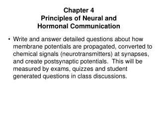
Chapter 4 Principles of Neural and Hormonal Communication • Write and answer detailed questions about how membrane potentials are propagated, converted to chemical signals (neurotransmitters) at synapses, and create postsynaptic potentials. This will be measured by exams, quizzes and student generated questions in class discussions.
Outline • Graded Potentials • Action Potentials • Synapses and integration • Intracellular communication • Signal Transduction • Hormonal Communication • Nervous vs. Endocrine System
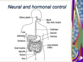
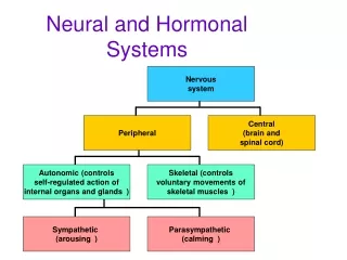
Communication is critical for the survival of the cells that compose the body. Two major regulatory systems of the body – nervous and endocrine - communicate with the cells/tissues/organs/systems they control. Neural communicationHormonal communication
Neural Communication • Nerve and muscle cells are excitable tissues. • Can alter their membrane permeabilities. • Permeability changes lead to membrane potential changes. • Potential changes act as electrical signals. • Electrical signals are necessary for normal nerve and muscle function.
Neural Communication +20 +10 Depolarization (decrease in potential; membrane less negative) 0 –10 Repolarization (return to resting potential after depolarization) –20 –30 Hyperpolarization (increase in potential; membrane more negative) Membrane potential (mV) –40 –50 –60 Resting potential –70 –80 –90 • Membrane electrical states • Polarization • Any state when the membrane potential is other than 0mV • Depolarization • Membrane becomes less polarized than at resting potential • Repolarization • Membrane returns to resting potential after having been depolarized • Hyperpolarization • Membrane becomes more polarized than at resting potential Time (msec) Fig. 4-1, p. 90
Voltage clamp • The technique allows an experimenter to "clamp" the cell potential at a chosen value. • This makes it possible to measure how much ionic current crosses a cell's membrane at any given voltage. • This is important because many of the ion channels in the membrane of a neuron are voltage gated ion channels, which open only when the membrane voltage is within a certain range. https://www.youtube.com/watch?v=Wd_gKJoo25Y Kenneth Cole[2] and George Marmount
Patch clamp • A patch-clamp microelectrode is a micropipette with a relatively large tip diameter. • The microelectrode is placed next to a cell, and gentle suction is applied through the microelectrode to draw a piece of the cell membrane (the 'patch') into the microelectrode tip; the glass tip forms a high resistance 'seal' with the cell membrane. • This can be used for studying the activity of the ion channels that are present in the patch of membrane. • If more suction is now applied, the small patch of membrane in the electrode tip can be displaced, leaving the electrode sealed to the rest of the cell. • This "whole-cell" mode allows very stable intracellular recording. https://www.youtube.com/watch?v=8LDO0hWWc0Q This technique was developed by Erwin Neher and Bert Sakmann who received the Nobel Prize in 1991.
Channels • Leak channels • Unregulated passage of ions • Gated channels • Voltage gated • Chemically gated • Mechanically gated • Thermally gated • These channels create and alter membrane potentials • Two kinds of potential change • Graded potentials • Serve as short-distance signals • Action potentials • Serve as long-distance signals
Graded Potential • Occurs in small, specialized region of excitable cell membranes • Magnitude of graded potential varies directly with the magnitude of the triggering event • Die out over short distances
Graded Potentials Examples of graded potentials: • Postsynaptic potentials • Receptor potentials • End-plate potentials • Pacemaker potentials • Slow-wave potentials
Action Potentials • Brief, rapid, large (100mV) changes in membrane potential during which potential actually reverses • Involves only a small portion of the total excitable cell membrane • Do not decrease in strength as they travel from their site of initiation throughout remainder of cell membrane
+70 +60 +50 +40 +30 +20 Action potential +10 0 –10 Membrane potential (mV) 20 –30 –40 –50 Threshold potential –60 –70 Resting potential –80 After hyperpolarization –90 Time (msec) 1 msec Slow depolarization to threshold Fig. 4-4, p. 94
VOLTAGE-GATED SODIUM CHANNEL VOLTAGE-GATED POTASSIUM CHANNEL Activation gate ECF Plasma membrane ICF Rapid opening triggered at threshold Slow closing triggered at threshold Delayed opening triggered at threshold Activation gate Inactivation gate (a) Closed but capable of opening (b) Open (activated) (c) Closed and not capable of opening (inactivated) (d) Closed (e) Open Fig. 4-5, p. 95
Action Potentials • When membrane reaches threshold potential • (-50 to-55mv) • Voltage-gated channels in the membrane undergo conformational changes • Flow of sodium ions into the ICF reverses the membrane potential from -70 mV to +30 mV • Flow of potassium ions into the ECF restores the membrane potential to the resting state
Action Potentials • Additional characteristics • Sodium channels open during depolarization by positive feedback. • When the sodium channels become inactive, the channels for potassium open. This repolarizes the membrane. • As the action potential develops at one point in the plasma membrane, it regenerates an identical action potential at the next point in the membrane. • Therefore, it travels along the plasma membrane undiminished.
4 Na+ channel closes and is inactivated (activation gate still open; inactivation gate closes) K+ channel opens (activation gate opens) Na+ channel reset to closed but capable of opening (activation gate closes; inactivation gate opens) K+ channel closes (activation gate closes) Na+ channel opens and is activated (activation gate opens; inactivation gate already open) Na+ in → rising phase 3 5 Membrane potential (mV) K+ out → falling phase K+ voltage-gated channel closed (activation gate closed) 2 ECF Threshold potential 6 1 8 Resting potential 7 Depolarizing triggering event ICF Time (msec) Na+ voltage-gated channel closed (activation gate closed; inactivation gate open) Fig. 4-7a, p. 96
Action Potentials The Na+/K+ pump gradually restores the concentration gradients disrupted by action potentials. • Sodium is pumped into the ECF • Potassium is pumped into the ICF • Refractory period keeps the action potential going in one direction and limits the Ap frequency • All or none • Frequency and line coding
Action Potentials • The Na+–K+ pump gradually restores the ions that moved during the action potential. • After an impulse has occurred in a patch of membrane, the membrane enters its refractory period. • It is impossible to re-stimulate the patch of membrane until it has recovered from its refractory period.
Refractory Periods • Absolute refractory period- period of time when a patch of membrane cannot be re-stimulated no matter how strong the stimulus. • Relative refractory period- period of time during which a patch of membrane can only be re-stimulated by a stronger than normal stimulus. • Refractory periods ensure the one-way propagation of action potentials.
New adjacent inactive area into which depolarization is spreading; will soon reach threshold Previous active area returned to resting potential New active area at peak of action potential “Backward” current flow does not reexcite previously active area because this area is in its refractory period “Forward” current flow excites new inactive area Direction of propagation of action potential Fig. 4-10, p. 101
Neuron • Once initiated, action potentials are conducted throughout a nerve fiber • Action potentials are propagated from the axon hillock to the axon terminals • Basic parts of neuron (nerve cell) • Cell body • Dendrites • Axon
Neuron • Cell body • Houses the nucleus and organelles • Dendrites • Project from cell body and increase surface area available for receiving signals from other nerve cells • Signal toward the cell body Dendrite and cell body serve as the neurons input zone.
Neuron • Axon • Nerve fiber • Single, elongated tubular extension that conducts action potentials away from the cell body • Conducting zone of the neuron • Collaterals • Side branches of axon • Axon hillock • First portion of the axon plus the region of the cell body fro m which the axon leaves • Neuron’s trigger zone • Axon terminals • Release chemical messengers that simultaneously influence other cells with which they come into close association • Output zone of the neuron
Action Potentials • Two types of propagation • Contiguous conduction • Conduction in unmyelinated fibers • Action potential spreads along every portion of the membrane • Saltatory conduction • Rapid conduction in myelinated fibers • Impulse jumps over sections of the fiber covered with insulating myelin
Contiguous Conduction Adjacent area that was brought to threshold by local current flow; now active at peak of action potential New adjacent inactive area into which depolarization is spreading; will soon reach threshold Previous active area returned to resting potential; no longer active; in refractory period Remainder of axon still at resting potential Fig. 4-9b, p. 100
Contiguous Conduction Adjacent inactive area into which depolarization is spreading; will soon reach threshold Active area at peak of action potential Remainder of axon still at resting potential Local current flow that depolarizes adjacent inactive area from resting potential to threshold potential Graded potential > threshold Direction of propagation of action potential Fig. 4-9a, p. 100
Saltatory Conduction Nodes of Ranvier Myelin sheath Myelin sheath Axon Axon of neuron Plasma membrane (a) Myelinated fiber Fig. 4-12a, p. 103
Myelin sheath Voltage-gated Na+ and K+ channels Axon Node of Ranvier Fig. 4-12d, p. 103
Saltatory Conduction • Propagates action potential faster than contiguous conduction because action potential does not have to be regenerated at myelinated section • Myelinated fibers conduct impulses about 50 times faster than unmyelinated fibers of comparable size • Myelin • Primarily composed of lipids • Formed by oligodendrocytes in CNS • Formed by Schwann cells in PNS
Adjacent inactive node into which depolarization is spreading; will soon reach threshold Active node at peak of action potential Remainder of nodes still at resting potential Local current flow that depolarizes adjacent inactive node from resting to threshold Direction of propagation of action potential Fig. 4-13, p. 104
Regeneration of Nerve Fibers • Regeneration of nerve fibers depends on its location • Schwann cells in PNS guide the regeneration of cut axons • Fibers in CNS myelinated by oligodendrocytes do not have regenerative ability • Oligodendrocytes inhibit regeneration of cut central axons
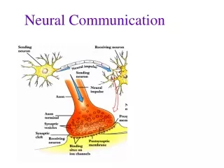
Synapses • Junction between two neurons • Primary means by which one neuron directly interacts with another neuron (muscle cells or glands as well) • Anatomy of a synapse • Presynaptic neuron – conducts action potential toward synapse • Synaptic knob – contains synaptic vesicles • Synaptic vesicles – stores neurotransmitter (carries signal across a synapse) • Postsynaptic neuron – neuron whose action potentials are propagated away from the synapse • Synaptic cleft – space between the presynaptic and postsynaptic neurons
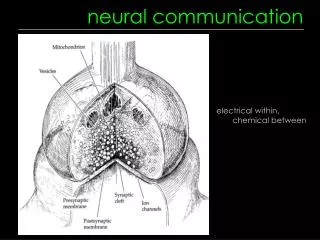
1 Axon of presynaptic neuron Synaptic knob (presynaptic axon terminal) 3 Voltage-gated Ca2+ channel Ca2+ Synaptic vesicle 2 Neuro- transmitter molecule Synaptic cleft 3 4 Subsynaptic membrane 5 4 Chemically gated receptor-channel for Na+, K+, or Cl– Postsynaptic neuron 5 Receptor for neurotransmitter Fig. 4-15, p. 108
Axon terminal of presynaptic neuron Dendrite of postsynaptic neuron Synaptic vesicles Synaptic cleft Fig. 4-15, p. 108
Neurotransmitters • Vary from synapse to synapse • Same neurotransmitter is always released at a particular synapse • Quickly removed from the synaptic cleft
Synapses Signal at synapse either excites or inhibits the postsynaptic neuron • Two types of synapses • Excitatory synapses • Inhibitory synapses • If binding of NT opens Na+ and K+ channels the result is a small depolarization called an excitatory post-synaptic potential (EPSP). • EPSP brings the cell closer to threshold. • If binding of NT opens either K+ or Cl– channels the result is a small hyperpolarization called an inhibitory post-synaptic potential (IPSP). • IPSP means cell less likely to reach threshold.
Synaptic Summation Cell body of postsynaptic neuron Axon hillock
Synaptic Summation • Multiple EPSP and IPSP’s from numerous synapses converge on one neuron. • These signals can cause different changes in the postsynaptic neuron • Cancellation • Spatial summation • Temporal summation
Excitatory presynaptic inputs (a) No summation (b) Temporal summation (c) Spatial summation (d) EPSP-IPSP cancellation +30 Membrane potential recorded 0 Postsynaptic membrane potential (mV) Threshold potential Resting potential –50 Postsynaptic cell –70 Inhibitory presynaptic input Time (msec) Fig. 4-17, p. 111
Presynaptic inputs Convergence of input (one cell is influenced by many others) Postsynaptic neuron Presynaptic inputs Divergence of output (one cell influences many others) Postsynaptic neurons Arrows indicate direction in which information is being conveyed. Fig. 4-20, p. 111
Synaptic Drug Interactions • Possible drug actions • Altering the synthesis, axonal transport, storage, or release of a neurotransmitter • Modifying neurotransmitter interaction with the postsynaptic receptor • Influencing neurotransmitter reuptake or destruction • Replacing a deficient neurotransmitter with a substitute transmitter
Examples of drugs that alter synaptic transmission • Cocaine • Blocks reuptake of neurotransmitter dopamine at presynaptic terminals • Strychnine • Competes with inhibitory neurotransmitter glycine at postsynaptic receptor site • Tetanus toxin • Prevents release of inhibitory neurotransmitter GABA, affecting skeletal muscles
DIRECT INTERCELLULAR COMMUNICATION Small molecules and ions (a) Gap junctions (b) Transient direct linkup of cells’ surface markers INDIRECT INTERCELLULAR COMMUNICATION VIA EXTRACELLULAR CHEMICAL MESSENGERS Secreting cell Local target cell Local target cell Electrical signal Secreting cell (neuron) Paracrine Neurotransmitter (c) Paracrine secretion (d) Neurotransmitter secretion Blood Secreting cell (endocrine cell) Blood Neurohormone Electrical signal Hormone Distant target cell Distant target cell Secreting cell (neuron) Nontarget cell (no receptors) Nontarget cell (no receptors) (e) Hormonal secretion (f) Neurohormone secretion Fig. 4-20, p. 117