Download
1 / 23
230 likes | 399 Vues
MICROBIOLOGY – ALCAMO. LECTURE: SPECIMEN PREPARATION AND STAINING . 1. INTRODUCTION. Why? --- MOs are small and transparent --- Cytoplasm of bacteria lacks color --- Stains enhance visibility. 2. WET SPECIMEN PREPARATIONS. ORGANISMS ARE NOT DRIED BEFORE HANDLING. WET MOUNT.

E N D
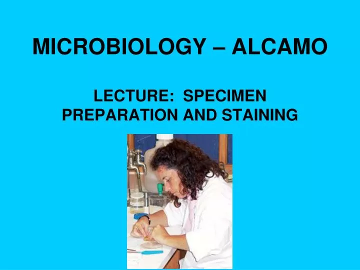
MICROBIOLOGY – ALCAMO LECTURE: SPECIMEN PREPARATION AND STAINING
1. INTRODUCTION Why? --- MOs are small and transparent --- Cytoplasm of bacteria lacks color --- Stains enhance visibility
2. WET SPECIMEN PREPARATIONS ORGANISMS ARE NOT DRIED BEFORE HANDLING
WET MOUNT • Quick and easy • Since no stain is used only large dense organisms are visible • TECHNIQUE: • Place drop of specimen on clean slide • Place cover slip over it
Simple Staining • Positively and negatively charged molecules are attracted to each other • MO’s cytoplasm has (-) charge • Basic stains have (+) charge • Crystal Violet • Methylene Blue • Therefore: Use (+) stains to color (-) MOs
NEGATIVE STAIN • Easy, fast, good for size evaluation • Stain is acidic and negatively charged: • Nigrosin (black dye) • Congo Red • Stains the background, not the MO • No need for chemicals and heat fixing • Cells appear less shriveled and distorted – more natural
TECHNIQUE: • PLACE DROP OF STAIN AT END OF SLIDE • DROP OF MOs ½ INCH BEFORE STAIN • WITH 2ND SLIDE HELD AT 45*, DRAW ACROSS MOs, THEN ACROSS STAIN • REVERSE DIRECTION, SMEAR FORWARD
3. DRY PREPARATIONS • MOs are dried and killed by “FIXING” • To flame quickly 3X Simple Differential
SIMPLE STAIN • One color dye only • EX: Crystal Violet, Methylene Blue • Easy, fast stain method with good results • TECHNIQUE: • Add the MO to slide • Air dry the MO • Fix the MO – Put through flame 3X • Flood with stain • Rinse with water • Dry for microscopic examination
DIFFERENTIAL STAIN • GRAM staining differentiates bacteria into 2 groups based on the differences in cell walls • Use two different colored dyes • All bacteria absorb the first stain color • But some lose the color when rinsed with alcohol and are stained with a 2nd color stain • Results are somewhat difficult and variable • Named for Christian Gram – Dutch physician
DIFFERENTIAL STAIN • GRAM (+) bacteria have peptidoglycan in their cell walls and retain the initial purple stain • GRAM (–) bacteria have more lipids in their cell wall and treatment with alcohol dissolves the lipids and the purple color leaks out • The GRAM (-) bacteria are now colorless, so a 2nd stain is needed to color these MO’s
DIFFERENTIAL STAIN • TECHNIQUE: • Stain with Crystal Violet (all MO’s are purple) • Cover with Gram’s iodine • Decolorize with alcohol • G+ stay purple • G- will lose the purple dye • Stain with Safranin dye (G- MO now appear red)
Gram (-) Gram (+)
Acid Fast Stain • Acid-fastness is a physical property of some bacteria referring to their resistance to de-colorization by acids during staining procedures • The high mycolic acid content of certain bacterial cell walls, like those of Mycobacteria, is responsible for the staining pattern of poor absorption followed by high retention
Acid Fast Stain • The most common staining technique used to identify acid-fast bacteria is the Ziehl-Neelsen stain, in which the acid fast bacilli are stained bright red and stand out clearly against a blue background.
SPECIAL STAINS • Involve special complicated methods not for amateurs • Used to observe special structures: • ENDOSPORES • FLAGELLA • CAPSULES