Download
1 / 45
470 likes | 753 Vues
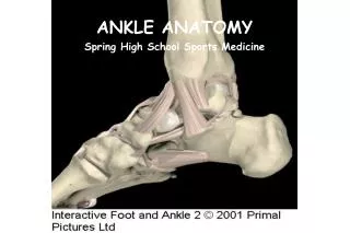
“Cartilage and Bone”. Cartilage. Embryo More prevalent than in adult Skeleton initially mostly cartilage Bone replaces cartilage in fetal and childhood periods. Location of cartilage in adults. External ear Nose “Articular” – covering the ends of most bones and movable joints

E N D
Cartilage • Embryo • More prevalent than in adult • Skeleton initially mostly cartilage • Bone replaces cartilage in fetal and childhood periods
Location of cartilage in adults • External ear • Nose • “Articular” – covering the ends of most bones and movable joints • “Costal” – connecting ribs to sternum • Larynx - voice box
Epiglottis – flap keeping food out of lungs • Cartilaginous rings holding open the air tubes of the respiratory system (trachea and bronchi) • Intervertebral discs • Pubic symphysis • Articular discs such as meniscus in knee joint
Remember the four basic types of tissue… • Epithelium • Connective tissue • Connective tissue proper • Cartilage • Bone • Blood • Muscle tissue • Nervous tissue
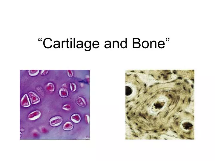
Cartilage is connective tissue • Cells called chondrocytes • Abundant extracellular matrix • Fibers: collagen & elastin • Jellylike ground substance of complex sugar molecules • 60-80% water (responsible for the resilience) • No nerves or vessels (hyaline cartilage)
Types of cartilage: 3 • Hyaline cartilage: flexible and resilient • Chondrocytes appear spherical • Lacuna – cavity in matrix holding chondrocyte • Collagen the only fiber • Elastic cartilage: highly bendable • Matrix with elastic as well as collagen fibers • Epiglottis, larynx and outer ear • Fibrocartilage: resists compression and tension • Rows of thick collagen fibers alternating with rows of chondrocytes (in matrix) • Knee menisci and annunulus fibrosis of intervertebral discs
Before we look at collagen pic… • Hyaline cartilage: flexible and resilient • Chondrocytes appear spherical • Lacuna – cavity in matrix holding chondrocyte • Collagen the only fiber • Elastic cartilage: highly bendable • Matrix with elastic as well as collagen fibers • Epiglottis and larynx • Fibrocartilage: resists compression and tension • Rows of thick collagen fibers alternating with rows of chondrocytes (in matrix) • Knee menisci and annulus fibrosis of intervertebral discs
Triple helix of collagen molecules form fibril Fibrils aggregate into collagen fibers
Growth of cartilage • Appositional • “Growth from outside” • Chrondroblasts in perichondrium (external covering of cartilage) secrete matrix • Interstitial • “Growth from within” • Chondrocytes within divide and secrete new matrix • Cartilage stops growing in late teens (chrondrocytes stop dividing) • Regenerates poorly in adults
Now about bones…like other connective tissue: cells separated by extracellular matrix with collagen but also mineral crystals Bone
Bones • Functions • Support • Movement: muscles attach by tendons and use bones as levers to move body • Protection • Skull – brain • Vertebrae – spinal cord • Rib cage – thoracic organs • Mineral storage • Calcium and phosphorus • Released as ions into blood as needed • Blood cell formation and energy storage • Bone marrow: red makes blood, yellow stores fat
Classification of bones by shape • Long bones • Short bones • Flat bones • Irregular bones • Pneumatized bones • Sesamoid bones (Short bones include sesmoid bones)
Gross anatomy of bones • Compact bone • Spongy (trabecular) bone • Blood vessels • Medullary cavity • Membranes • Periosteum • Endosteum
Flat bones • Spongy bone is called diploe when its in flat bones • Have bone marrow but no marrow cavity
Long bones • Tubular diaphysis or shaft • Epiphyses at the ends: covered with “articular” (=joint) cartilage • Epiphyseal line in adults • Kids: epiphyseal growth plate (disc of hyaline cartilage that grows to lengthen the bone) • Blood vessels • Nutrient arteries and veins through nutrient foramen
Periosteum • Connective tissue membrane • Covers entire outer surface of bone except at epiphyses • Two sublayers • 1. Outer fibrous layer of dense irregular connective tissue • 2. Inner (deep) cellular osteogenic layer on the compact bone containing osteoprogenitor cells (stem cells that give rise to osteoblasts) • Osteoblasts: bone depositing cells • Also osteoclasts: bone destroying cells (from the white blood cell line) • Secured to bone by perforating fibers (Sharpey’s fibers) Endosteum • Covers the internal bone surfaces • Is also osteogenic
Bone markings • Projections that are the attachments sites for muscles and ligaments • Surfaces that form joints • Depressions and openings Learn them using: Marieb lab book p 101, Table 8.1, Bone Markings or Martini p 128, Table 5.1, Common Bone Marking Terminology (next slide)
Martini p 128, Table 5.1, Common Bone Marking Terminology (for figure see next slide)
Compact bone • Osteons: pillars • Lamellae: concentric tubes • Haversian canals • Osteocytes
Isolated osteon: • Nutrients diffuse from vessels in central canal • Alternating direction of collagen fibers increases resistance to twisting forces
Spongy bone • Layers of lamellae and osteocytes • Seem to align along stress lines
Chemical composition of bones • Cells, matrix of collagen fibers and ground substance (organic: 35%) • Contribute to the flexibility and tensile strength • Mineral crystals (inorganic: 65%) • Primarily calcium phosphate • Lie in and around the collagen fibrils in extracellular matrix • Contribute to bone hardness • Small amount of water
Bone development • Osteogenesis: “formation of bone” • From osteoblasts • Bone tissue first appears in week 8 (embryo) • Ossification: “to turn into bone” • Intramembranous ossification (also called “dermal” since occurs deep in dermis): forms directly from mesenchyme (not modeled first in cartilage) • Most skull bones except a few at base • Clavicles (collar bones) • Sesamoid bones (like the patella) • Endochondral ossification: modeled in hyaline cartilage then replaced by bone tissue • All the rest of the bones
Remember the three germ tissues… • Ectoderm- epithelial • Endoderm- epithelial • Mesoderm is a mesenchyme tissue • Mesenchyme cells are star shaped and do not attach to one another, therefore migrate freely • From the last slide: Intramembranous ossification: forms directly from mesenchyme (not modeled first in cartilage) • Most skull bones except a few at base • Clavicles (collar bones) • Sesmoid bones (like the patella)
Intramembranous ossification (osteoid is the organic part)
Endochondral ossification • Modeled in hyaline cartilage, called cartilage model • Gradually replaced by bone: begins late in second month of development • Perichondrium is invaded by vessels and becomes periosteum • Osteoblasts in periosteum lay down collar of bone around diaphysis • Calcification in center of diaphysis • Primary ossification centers • Secondary ossification in epiphyses • Epiphyseal growth plates close at end of adolescence • Diaphysis and epiphysis fuse • No more bone lengthening See next slide
Endochondral ossification Stages 1-3 during fetal week 9 through 9th month Stage 5 is process of long bone growth during childhood & adolescence Stage 4 is just before birth
Organization of cartilage within the epiphyseal plate of a growing long bone
Epiphyseal growth plates in child, left, and lines in adult, right (see arrows)
Factors regulating bone growth • Vitamin D: increases calcium from gut • Parathyroid hormone (PTH): increases blood calcium (some of this comes out of bone) • Calcitonin: decreases blood calcium (opposes PTH) • Growth hormone & thyroid hormone: modulate bone growth • Sex hormones: growth spurt at adolescense and closure of epiphyses
Bone remodeling • Osteoclasts • Bone resorption • Osteoblasts • Bone deposition • Triggers • Hormonal: parathyroid hormone • Mechanical stress • Osteocytes are transformed osteoblasts
Terms (examples) • chondro refers to cartilage • chondrocyte • endochondral • perichondrium • osteo refers to bone • osteogenesis • osteocyte • periostium • blast refers to precursor cell or one that produces something • osteoblast • cyte refers to cell • osteocyte
Repair of bone fractures (breaks) • Simple and compound fractures • Closed and open reduction
Disorders of cartilage and bone • Defective collagen • Numerous genetic disorders • eg. Osteogenesis imperfecta (brittle bones) – AD (autosomal dominant) • eg. Ehlers-Danlos (rubber man) • Defective endochondral ossification • eg. Achondroplasia (short –limb dwarfism) - AD • Inadequate calcification (requires calcium and vitamin D) • Osteomalacia (soft bones) in adults • Rickets in children Note: “AD” here means autosomal dominant inheritance
(continued) • Pagets disease – excessive turnover, abnormal bone • Osteosarcoma – bone cancer, affecting children primarily • Osteoporosis – usually age related, esp. females • Low bone mass and increased fractures • Resorption outpaces bone deposition
Normal bone Osteoporotic bone