Download
1 / 41
600 likes | 1.24k Vues
The Digestive System- the digestion of food begins in the mouth. What does it do?. The digestive system consists of several organs that work together to: ingest food Breakdown food into smaller molecules, so that the nutrients that can cross the cell membrane absorb nutrients

E N D
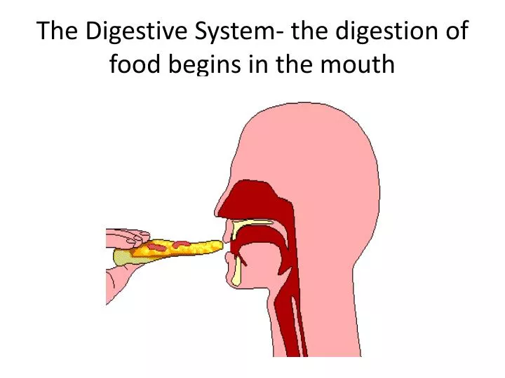
The Digestive System- the digestion of food begins in the mouth
What does it do? • The digestive system consists of several organs that work together to: • ingest food • Breakdown food into smaller molecules, so that the nutrients that can cross the cell membrane • absorb nutrients • Eliminate indigestible remains (wastes) from the body
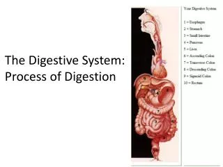
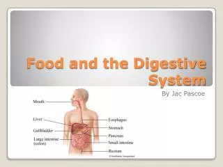
Passageway of food as it enters the digestive system • Mouth, pharynx, esophagus, stomach, small intestine, large intestine, rectum, anal canal • Organs involved in digestion that food never pass through: liver, gall bladder, pancreas
There are two types of digestion Mechanical Digestion Chemical Digestion Digestive enzymes break down macromolecules into small organic molecules that can be absorbed by cells • Large pieces of food become smaller pieces, getting it ready for chemical digestion • Begins with the teeth chewing food in the mouth • Churning and mixing of food in stomach
Some Digestive Enzymes • Carbohydrates are first broken down in the mouth chemically by the enzyme in saliva called amylase • Proteins arefirst broken down in the stomach by the enzyme, pepsin • Lipase first begins the break down of fats in the small intestine
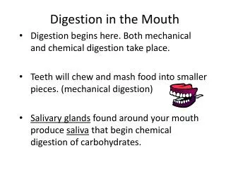
Mouth • Digestion begins here!! • Sensory receptors on tongue, taste buds, are activated by the presence of food which causes a nerve impulse to travel to the brain via the cranial nerve • Teeth are involved in mechanical digestion, allowing us to chew food to pieces small enough for swallowing • Three pairs of salivary gland in the mouth secrete saliva which contains an enzyme called, amylase, which begins the process of chemically digesting starches, which are carbohydrates
The Three pairs of salivary glands in the mouth secrete saliva which moisten food and breaks down carbohydrates with its amylase enzyme
The Pharynx (throat) • The region that receives air from the nasal cavity and food from the mouth • Swallowing, which occurs in the pharynx is a reflex action performed automatically, without conscious thought • When you swallow, a flap of tissue called the epiglottis, closes over the trachea so that food does not go down the airway and cause a person to choke • For this reason, we do not breathe when we swallow
The green pathway represents what happens when you swallow- red is when you are not swallowing
Esophagus • A muscular tube that passes from the pharynx to the stomach that opens to receive the bolus (chewed up food) • Waves of muscular contraction called peristalsis pushes food along the digestive tract • Peristalsis begins in the esophagus and continues in the stomach and intestines • Sphincters are muscles that encircle tubes that act as valves. There is a sphincter at the meeting of the esophagus and stomach • Relaxation of the sphincter allows the bolus to enters, while contraction prevents the acidic content from the stomach from backing up into the esophagus
Peristalsis of the esophagus wall passes food along to the stomach
Did you know? • Heartburn feels like a burning pain rising up in the throat. It occurs when some of the contents in the stomach escape into the esophagus • When vomiting occurs, a contraction of the abdominal muscles and diaphragm propels food upward through the esophagus • Did you know that if you stand on your head, you can eat and the food will still go down (or in this case- up ) your esophagus and enter your stomach? That is how powerful peristalsis action is!
The Stomach • A thick-muscular walled, J-shaped organ that lies on the left side of the liver • Length of stomach remains about 10 in regardless of amount of food, but it can expand in diameter, holding up to a 1 to 1.5 liters of food! • The stomach receives food from the esophagus, stores it, mixes it with its digestive juices, and breaks down proteins into smaller pieces
Stomach • The term gastric always refers to the stomach, so what does a gastroenterologist treat? Diseases of the…. • The gastric glands produce gastric juice which contains pepsinogen, HCI, and mucus • Chief cells secrete pepsinogen, which becomes the enzyme pepsin (breaks down protein)when exposed to hydrochloric acid • HCI is released by parietal cells- the HCI causes the stomach to have a pH of about 2 and is beneficial because it kills most of the bacteria present in food and helps break down food . Stomach cells are replaced through mitosis every 2-3 days because of the harsh acidic enviroment
More on the Stomach • Alcohol is absorbed in the stomach, but food and other substances are not. They are absorbed later in the small intestine • Normally, the stomach empties in about 2-4 hours • When food leaves the stomach, it is a thick, soupy liquid called chyme. • Chyme enters the small intestine by way of the pyloric sphincter, which acts as a valve between the stomach and the small intestine
Hydrochloric Acid • HCl simply breaks down the connective tissue of meat and activates pepsin • The wall of the stomach is protected by a thick mucous layer which secretes mucus • If HCl penetrates the mucous layer, the wall can begin to break down and an ulcer results • An ulcer is an open sore in the wall caused by the gradual destruction of tissue. Most are due to a bacterial infection (Helicobacter pylori) that impairs the ability of mucous cells to secrete mucus • Stress and spicy foods don’t cause ulcers, but they can aggravate the condition
Hydrochloric Acid (HCl) • Kills bacteria and other living cells in food • Causes pepsinogen to be converted to its active form pepsin • Denatures proteins which makes it easier for enzymes to act on them • Causes the stomach to have a low pH which is optimum for the pepsin enzyme
You Be The Doctor! How do you think a doctor might treat a patient with an ulcer caused by H. Pylori bacteria? Reduce stomach acid Kill the bacteria with antibiotics Protect the stomach lining
The Small Intestine • The small intestine is about 20 feet in length • Ducts from the liver and pancreas join to form one duct that enters the duodenum. The small intestine receives bile from liver and pancreatic juices from the pancreas • Bile, a yellowish green, thick mucus produced by the liver emulsifies (loosens up) fat, causing fat droplets to disperse in water, then lipase can easily break down fats • The small intestine has a slightly basic pH because the pancreatic juice contains sodium bicarbonate (NaHCO3) which neutralizes chyme
Villi • The wall of the small intestine contain tiny, finger-like projections called villi, which give the wall a soft velvety appearance • Each villus has an outer layer of columnar epithelial cells, and each of these cells has thousands of microscopic extensions called microvilli • The microvilli greatly increase the surface area of the villus for the absorption of nutrients • In each villus there are blood capillaries and lymph vessels • After nutrients are absorbed, they are carried to all the cells of the body by the bloodstream
The Small Intestine • The pancreas secretes digestive juices with enzymes into the small intestine to complete the digestion of food • The small intestine is where the absorption of most nutrients takes place
The surface area of the small intestine is approximately the size of a tennis court! Imagine that.
Pancreas • The pancreas is a gland that lies deep in the abdominal cavity, resting just beneath the stomach • Most pancreatic cells produce pancreatic juice, which contains sodium bicarbonate and a mix of digestive enzymes that are secreted in the small intestine to finish the break down of nutrients: • Pancreatic amylase completes the digestion of starch • Trypsincompletesthe digestion of proteins • Lipase completes the digestion of fats
Notice the pancreas and liver both have ducts emptying into the small intestine; the pancreas secretes digestive juices into the small intestine to finish the break down of carbohydrates, fats, and protein and bile loosens up fat
The Liver • Produces bile • It detoxifies blood by removing and metabolizing poisonous substances • Stores sugar, iron (Fe) and the fat soluble vitamins, A, D, K, E, and B12 • Helps regulate blood cholesterol, converting some to bile salts • It maintains the blood glucose levels • Removes bilirubin, a breakdown product of hemoglobin from the blood and excretes it in bile • Produces urea after breaking down amino acids • Makes clotting factors to stop excess bleeding
The Liver The largest organ in the body. It is a complex organ that you cannot live without! Drinking too much alcohol and a eating a diet heavy in fat can damage liver tissue over time. Question for you to research: Do you think you can you live without a gall bladder?
Gall bladder • The gall bladder is attached to the surface of the liver • It stores excess bile produced by the liver and when needed, it releases bile into the duodenum • The passage of the stones may block the common bile duct and cause jaundice, a yellowish tint of the whites of the eyes and skin. Jaundice is caused by abnormal amounts of bilirubin in the blood. At this point, the gall bladder must be removed
The Large Intestine • The large intestine includes the cecum, the colon, the rectum, and the anus • It is larger in diameter than the small intestine, but shorter in length. It is about 41/2 feet long • Water, salts, and some vitamins are absorbed here. • It also stores indigestible materials until it is eliminated at the anus
Large Intestine • Inside the large intestine are bacteria. Over 99% of the intestinal bacteria are anaerobes (bacteria that die in the presence of oxygen) • The bacteria are helpful in that they break down indigestible material, and produce some vitamins that are body uses (they also produce gas, or as you know it as- farts) So the next time you think all bacteria are bad…think again. Some are actually beneficial to us…
Problems with the Large Intestine • Two common everyday complaints associated with the large intestine are diarrhea and constipation • The major causes of diarrhea are infection of the lower intestinal tract. In the case of infection, such as food poisoning, the intestinal wall becomes irritated, and peristalsis increases. Water is not absorbed, and diarrhea results • The major causes of constipation, dry and hard feces, is a lack of water or fiber in the diet
Gastric Bypass Surgery This weight loss surgery is not for everyone! Here’s what’s involved: The surgeon creates a small pouch at the top of your stomach and adds a bypass around a segment of the stomach and the small intestine. The surgeon staples the stomach at the top, sealing it off from the rest of the stomach. The resulting pouch is about the size of a walnut and can hold only and ounce of food,. Then the surgeon cuts part of the small intestine and sews part of it to the pouch. Food enters the small intestine directly, limiting your ability to absorb calories
Cirrhosis of the Liver • Chronic disease of the liver where the liver becomes fatty, and then the liver tissue is replaced by fibrous scar tissue • This disease is often seen in alcoholics, due to malnutrition and to the excessive amounts of alcohol (a toxin) the liver is forced to break down • The liver has amazing regeneration powers and can recover if the rate of regeneration exceeds the rate of damage • During liver failure, however, there may not be enough time for it to heal itself, and liver transplant might be only option
Hepatitis • Inflammation of the liver caused by a virus; three common types: • Hepatitis A- usually acquired from sewage-contaminated drinking water; common in developing countries • Hepatitis B- usually spread through sexual contact, but also blood transfusions (prior to 1991) and infected needles; can be spread from mother to child • Hepatitis C- usually acquired by coming in direct contact with infected blood; likely to lead to chronic hepatitis, liver cancer and death
Tips: How to Take Care of Your Liver • Wash your hands thoroughly after using the washroom (A) • Eat a sensible diet with plenty of fruits and vegetables (washed and clean) (A) • If traveling to undeveloped countries- drink only bottled water and consider a hepatitis A vaccination (A) • Avoid unsafe sex practices (B and C) • Do not share needles, razors, tooth brushes, etc (B and C) • Keep alcohol intake very moderate, or abstain (C and cirrhosis) • Also a vaccine now for Hepatitis B
Gall Stones • What is it? • Gall stones are tiny, pebble-like stones that develop in the gall bladder • They form when the bile harden to form stone-like