Download
1 / 24
240 likes | 506 Vues
COMPLEX LIPID METABOLISM - I 10/01/07. LEARNING OBJECTIVES 1) To identify the basic structure of phospholipids and to be able to distinguish the two classes, phosphoglycerides and sphingomyelins 2) To be able to name and recognize the major phosphoglycerides

E N D
COMPLEX LIPID METABOLISM - I 10/01/07 • LEARNING OBJECTIVES • 1) To identify the basic structure of phospholipids and to be able to distinguish the two classes, phosphoglycerides and sphingomyelins • 2) To be able to name and recognize the major phosphoglycerides • To differentiate how phosphoglycerides are synthesized, and which of the building blocks contain the high energy bond • To summarize the way in which sphingomyelin is synthesized • To explain how phosphoglycerides and sphingomyelin are degraded
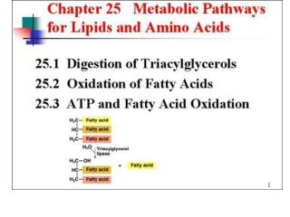
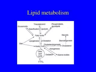
II. INTRODUCTION TO PHOSPHOLIPIDS A) Polar, ionic compounds (Fig. 17.1A) B) Contain an alcohol (diacylglycerol or sphingosine) and a phosphodiester linkage III. STRUCTURE A) Two Classes 1) glycerophospholipids (phosphoglycerides) – glycerol backbone; function in membrane structure, anchoring proteins in the cell membrane, signal transduction across membranes, bile composition, lipoprotein particles 2) sphingomyelins - sphingosine backbone - primarily in membrane structure
B) Phosphoglycerides (Fig. 17.1) 1) glycerol backbone 2) phosphate esterified at the three position 3) fatty acids esterified at the one and two positions [1) + 2) + 3) = phosphatidic acid (PA)] (Fig. 17.1B) – simplest phosphoglyceride 4) an alcohol component esterified on the phosphate 5) common phosphoglycerides: (Fig. 17.1A) PA + serine = phosphatidylserine (PS) PA + ethanolamine = phosphatidylethanolamine (PE, cephalin) PA + choline = phosphatidylcholine (PC, lecithin) PA + glycerol = phosphatidylglycerol (PG) PA + inositol = phosphatidylinositol (PI)
6) cardiolipin (Fig. 17.2) - important inner mitochondrial membrane component = two PA’s esterified to 1 and 3 position of another glycerol. The only phosphoglyceride that is antigenic. 7) plasmalogens (Fig. 17.3) - fatty acid at carbon 1 position is an ether instead of an ester linkage a) three classes - phosphatidalcholines, phosphatidal- ethanolamines, and phosphatidalserines b) myelin - has much ethanolamine plasmalogen c) heart muscle - has much choline plasmalogen d) platelet-activating factor (PAF) – strong biological mediator; causes platelet activation; increases pulmonary and airway edema in lungs; mediates hypersensitivity, acute inflammatory reactions, and anaphylactic shock; stimulates neutrophils and macrophages to produce superoxide radicals; binds to membrane receptors
C) Sphingomyelins (Fig. 17.4) 1) backbone is sphingosine 2) fatty acid forms amide linkage with amino group of sphingosine = a ceramide (this is also a precursor for glycolipids) 3) esterification of carbon 1 of ceramide by phosphorylcholine = sphingomyelin (important component of myelin in nerves)
IV. PHOSPHOGLYCERIDE SYNTHESIS (Fig. 17.5) - two methods A) Donation of phosphatidic acid from CDP~diacylglycerol to an alcohol B) Donation of phosphomonoester of the alcohol from the CDP~alcohol to 1,2-diacylglycerol C) CDP-derivative is “activated” (has a high energy bond) D) Synthesis occurs in smooth ER and goes through the Golgi apparatus to membranes or cell exterior (exocytosis) E) Phosphatidic acid synthesis (see Fig 16.14); occurs in all tissues except rbc’s
F) Phosphatidylethanolamine (PE) and phosphatidylcholine (PC) synthesis 1) ethanolamine and choline obtained from the diet or from phospholipid turnover; phosphatidylcholine can be synthesized de novo from PS 2) preexisiting - phosphorylation of ethanolamine or choline; activation to CDP-ethanolamine or CDP-choline; ethanolamine phosphate or choline phosphate is transferred to diacylglycerol (salvage pathway)
a) choline reutilization - de novo choline synthesis needs three methyl groups from methionine (Fig. 17.6, bottom). Because methionine (an essential amino acid) is often deficient in the diet, choline reutilization prevents possible choline deficiency b) lung surfactant – dipalmitoyl- phophatidylcholine (DPPC) is an important lung surfactant; hyaline membrane disease (respiratory distress syndrome, RDS) in premature infants = low lung surfactant production. Glucocorticoids (stimulate DPPC synthesis) can be given to the mother prior to delivery to reduce the possibility of RDS in the premature infant. Immune suppressive medication and chemotherapy – adult RDS 3) de novo synthesis of PC from PS (Fig. 17.6) - PS is decarboxylated and then methylated three times G) Phosphatidylserine (PS) (Fig. 17.6, top) – calcium- activated “base exchange” – the ethanolamine of PE is exchanged with free serine
H) Phosphatidylinositol (PI) 1) inositol + CDP-diacylglycerol PI and CMP (Fig. 17.5) 2) signal transduction - PI becomes phosphorylated (for example, to PIP2; Fig. 17.7).
When cells are treated with hormones, neurotransmitters, and growth factors, the degradation of PIP2 by phospholipase C is stimulated. This yields inositol-1,4,5-trisphosphate (IP3; mobilizes intracellular calcium) and diacylglycerol (which activates protein kinase C) (Figs. 17.7 and 17.8). DAG IP3
3) membrane protein anchoring (Fig. 17.9) – important for anchoring of alkaline phosphatase, acetylcholine esterase, and lipoprotein lipase 4) A deficiency in glycosyl phosphatidylinositol (GPI) in hematopoietic cells causes a hemolytic disease, paroxysmal nocturnal hemoglobinuria. 5) phospholipase C cleavage of protein and inositol phosphate leaves diacylglycerol (which activates protein kinase C) I) Phosphatidylglycerol (PG) – mitochondrial membranes; precursor of cardiolipin; made from CDP-diacylglycerol and glycerol-P (Fig. 17.5) J) Cardiolipin (diphosphatidylglycerol) – diacylglycerolphosphate transferred from CDP-diacylglycerol to PG (Fig. 17.5)
V. SPHINGOMYELIN SYNTHESIS (Fig. 17.10) A) palmitoyl CoA and serine are used as substrates. A series of reactions that include decarboxylation, loss of CoA, reduction by NADPH + H+ and oxidation by FAD results in the synthesis of sphingosine. B) An amide bond is formed between sphingosine and a fatty acid (using fatty acyl CoA) to produce a ceramide. C) Using phosphatidylcholine, choline phosphate is transferred to the ceramide to form a sphingomyelin.
VI. PHOSPHOLIPID DEGRADATION (Fig. 17.11) A) Degradation of phosphoglycerides - various phospholipases cleave the phosphodiester bonds at specific sites 1) phospholipase A1 - removes fatty acid at position 1 2) phospholipase A2 - removes fatty acid at position 2; releases arachidonic acid for prostaglandin, leukotriene, and thromboxane synthesis; high in pancreatic secretion, activated by trypsin, and requires bile salts for activity; inhibited by glucocorticoids 3) phospholipase C - in liver lysosomes; plays role in producing second messengers in the PIP2 pathway; leaves free hydroxyl at position 3 of glycerol 4) phospholipase D - removes alcohol from the phosphoglyceride, leaving phosphatidic acid
B) Degradation of sphingomyelin (Fig. 17.12) 1) degraded by sphingomyelinase - lysosomal enzyme; removes phosphorylcholine to yield a ceramide. Niemann-Pick disease – sphingomyelinase deficiency. Severe form (Type A) has large accumulation of sphingomyelin and phosphatidylcholine in liver and spleen (both very enlarged) (Fig. 17.13). Results in severe mental retardation and death in early adulthood. “High” in Ashkenazi Jews. 2) ceramidase - cleaves ceramide into sphingosine and a free fatty acid.
COMPLEX LIPID METABOLISM - II 10/01/07 • I. LEARNING OBJECTIVES • To identify the structure of glycosphingolipids and to be able to distinguish the two classes, neutral glycosphingolipids and acidic • glycosphingolipids • 2) To be able to name and recognize the major gangliosides and sulfatides • To describe how glycosphingolipids are synthesized • 4) To summarize how glycosphingolipids are degraded • 5) To list the various sphingolipidoses and the enzymes that are responsible for the defect.
II. INTRODUCTION TO GLYCOLIPIDS A) Because they are derivatives of ceramide = “glycosphingolipids” B) Numerous functions 1) membrane component (particularly outer surface of plasma membrane; interact with the extracellular environment) 2) very high in nerve tissue 3) roles in cellular interactions, growth, development 4) blood group, embryonic, and tumor antigens 5) receptors for cholera and diphtheria toxins and for viruses 6) genetic disorders in their metabolism cause severe neurological and developmental problems
III. STRUCTURE A) General features - contain no phosphate and polar head group is comprised of mono- or oligosaccharides attached to ceramide by an O-glycosidic bond B) Neutral glycosphingolipids 1) cerebroside (Fig. 17.14) (found primarily in brain and peripheral nervous tissue; high concentrations in myelin sheaths) – contains monosaccharide unit only a) galactocerebroside (most common in membranes) contains galactose: Cer-Gal b) glucocerebroside (intermediate in glycosphingolipid synthesis) contains glucose: Cer-Glc 2) ceramide oligosaccharides (globosides; additional sugars are added to a glucocerebroside) – includes: a) lactosylceramide: Cer-Glc-Gal b) Forssman antigen: Cer-Glc-Gal-Gal-GalNac-GalNac* *GalNac = N-acetylgalactosamine
C) Acidic glycosphingolipids – negatively charged at physiological pH; due to either N-acetylneuraminic acid (NANA) (sialic acid) in gangliosides or sulfate in sulfatides 1) gangliosides (Fig. 17.15) - primarily in ganglion cells of CNS (nerve endings); derivatives of ceramide oligosaccharides. Naming includes “G” for ganglioside; followed by letter that designates how many NANA molecules are present (M = mono- = 1; D = di- = 2; T = tri- = 3; Q = quatro- = 4); additional letters or numbers in a subscript refer to a convention for the sequence of carbohydrate attachment to the ceramide (e.g., GM2) 2) sulfatides (sulfoglycosphingolipids) – cerebrosides that contain a sulfated galactosyl residue. Found mostly in nervous tissue.
IV. GLYCOSPHINGOLIPID SYNTHESIS A) Mechanism (Fig. 17.18) - sequential addition of sugar (glycosyl) residues. Sugar nucleotide donors (highenergybond) used, similar to glycoprotein synthesis. Occurs in Golgi and smooth endoplasmic reticulum. B) Enzymes - glycosyl transferases that are specific for a nucleotide sugar and an acceptor. Some enzymes are involved in both glycoprotein and glycosphingolipid synthesis. X
C) Sulfate group addition - added to galactocerebrosides by transfer from 3’ phosphoadenosine-5’-phosphosulfate (PAPS). (Fig. 17.16) Sulfate is added to the 3’ hydroxyl group of galactose by sulfotransferase (Fig. 17.17). High Energy Bond
V. GLYCOSPHINGOLIPID DEGRADATION A) Internalized by endocytosis B) Degraded by lysosomal enzymes after fusion of endocytic vesicles with lysosomes C) Enzymes involved include: a-galactosidase, b-galactosidase, b-glucosidase, neuraminidase (sialidase), hexosaminidase, sphingomyelinase, sulfatase (sulfateesterase), and ceramidase. Remove residues sequentially such that the last one added during synthesis is the first one removed during degradation. Defects in the enzymes lead to a large spectrum of diseases called lipid storage diseases or sphingolipidoses. Have accumulation of undigested glycosphingolipids within cells (Fig. 17.19)
VI. SPHINGOLIPIDOSES (Fig. 17.20) A) Synthesis and degradation usually balanced B) Degradation (hydrolysis) occurs in lysosomes C) Deficiency in hydrolase causes lipid accumulation in the lysosome – Almost all show neurological impairment and most are fatal in early life. E) Except for Fabry Disease (X-linked) they are autosomal recessive. D) Diagnosis - assay enzyme activity or accumulated lipid in tissue biopsies, cultured fibroblasts, peripheral blood leukocytes, plasma, and/or amniotic fluid. E) Very rare in the general population. Gaucher and Tay-Sachs – Ashkenazi Jews
H) Effects of Eicosanoids (Fig. 17-25) – KNOW ALL OF THESE!! 1) many varied effects 2) Tromboxane A2 (TXA2) – promotes blood clots by causing platelet aggregation and vasoconstriction. Prostacyclin (PGI2) inhibits platelet aggregation and causes vasodilation, and inhibits thrombogenesis, by acting on nucleated endothelial cells. Aspirin blocks both by covalently attaching to COX-1 and inactivating it(Fig. 17-24). But endothelial cells can synthesize more COX-1 and make PGI2, while anucleated platelets cannot synthesize more TXA2. Basis for low-dose aspirin therapy for lowering stroke and heart attack risk by lowering thrombi formation. 3) COX-2 inhibitors may cause increased risk of heart attack and stroke (Vioxx recall).