Download
1 / 57
600 likes | 791 Vues
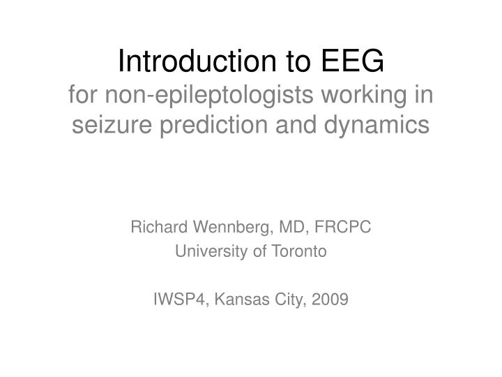
Introduction to EEG for non-epileptologists working in seizure prediction and dynamics. Richard Wennberg, MD, FRCPC University of Toronto IWSP4, Kansas City, 2009. 1. EEG source. cortical pyramidal cells voltage fluctuations in space/time summated EPSPs/IPSPs

E N D
Introduction to EEG for non-epileptologists working in seizure prediction and dynamics Richard Wennberg, MD, FRCPC University of Toronto IWSP4, Kansas City, 2009
1. EEG source • cortical pyramidal cells • voltage fluctuations in space/time • summated EPSPs/IPSPs • dependent on neural “synchrony”
2. EEG oscillations • Normal • (alpha, beta, mu, gamma, sleep spindles/delta) • generated in cortex • varying degrees of thalamocortical interdependence • Abnormal • (seizures, burst-suppression)
3. EEG sharp transients • Normal • Vertex sharp waves, positive occipital sharp transients of sleep (POSTS), benign epileptiform transients of sleep (BETS) or small sharp spikes; eye blinks, EKG, EMG • Abnormal • Epileptiform spikes, polyspikes, spike and waves, sharp waves, sharp and slow waves • Periodic complexes (lateralized and generalized), triphasic waves
Pedley and Traub. In: Daly and Pedley, eds. Current Practice of Clinical EEG, 1990
Pedley and Traub. In: Daly and Pedley, eds. Current Practice of Clinical EEG, 1990
Steriade. In: Niedermeyer and Lopes da Silva, eds. Electroencephalography, 1993
Subject awake, resting. Normal posterior alpha rhythm disappears with eye opening (*). High frequency activity at end of figure after eye opening is muscle artifact. Anterior-posterior bipolar montage.LFF 0.5 Hz, HFF 70 Hz, this and all other figures.
Stage II sleep. K-complex (*); Sleep spindles (**). Anterior-posterior bipolar montage.
Burst of generalized 3Hz spike and wave activity (*). Primary generalized epilepsy. Anterior-posterior bipolar montage.
Generalized, bilaterally synchronous, 3 Hz spike and wave activity in a different patient with primary generalized epilepsy. Referential montage; reference = linked ears.
Primary generalized epilepsy: spike and wave burstsJuvenile myoclonic epilepsy Generalized, bilaterally synchronous bursts of spike and wave activity in another patient with primary generalized epilepsy, subtype juvenile myoclonic epilepsy. Referential montage; reference = linked ears.
Primary generalized epilepsy: transition to tonic-clonic seizureJuvenile myoclonic epilepsy In this condition, bursts of spike and wave activity increase in frequency in the morning hours after awakening in a true “pre-ictal period” that may – or may not – result in a transition to a generalized tonic-clonic seizure.
Primary generalized epilepsy: transition to tonic-clonic seizureHigh amplitude “hypersynchrony” Same seizure transition as previous figure, shown here at slower sweep speed.
Bilateral temporal lobe (“focal”, “partial”) interictal epileptiform activity. Independent sharp and slow wave complexes over right (*) and left (**) anterior-mid temporal regions. Temporal lobe epilepsy. Anterior-posterior bipolar montage.
Ictal EEG showing focal rhythmic seizure pattern localized to right temporal region (“equipotentiality” at F8-T4). Temporal lobe epilepsy. Anterior-posterior bipolar montage.
Patient with bilateral hippocampal sclerosis, global developmental delay, medically-refractory complex partial seizures Interictal EEG during drowsiness in a different patient showing unilateral right anterior temporal lobe spikes (“phase reversing” at Zg2, F8, F10)
Patient with bilateral hippocampal sclerosis, global developmental delay, medically-refractory complex partial seizures Ictal EEG showing unilateral right temporal lobe seizure (with “equipotentiality” at Zg2-T4, F8-T4, F10-T10) (note different sensitivity and time scale compared with preceding, interictal EEG figure from same patient)
Why should the non-epileptologist care about artifacts and reference electrodes? Two examples • EEG studies of beta and gamma oscillations in cognition would appear to have been analyzing mainly muscle artifact • Whitham et al. Clin Neurophysiol 2008;119:1166-75 and Clin Neurophysiol 2007;118;1877-88 • The need for a reference electrode in EEG affects phase synchronization studies; resulting amplitude variations influence the phase locking analyses • Guevara et al. Neuroinformatics 2005;3:301-13
Eye blink, horizontal eye movements, frontalis and temporalis EMG, lateral rectus EMG, pulse artifacts. Combined circular and anterior-posterior bipolar montage.
Eye blink, horizontal eye movements, frontalis and temporalis EMG, lateral rectus EMG, pulse artifacts. Referential montage; reference = Pz.
Eye blink, horizontal eye movements, frontalis and temporalis EMG, lateral rectus EMG, pulse artifacts. Referential montage; reference = Fz.
Eye blink, horizontal eye movements, frontalis and temporalis EMG, lateral rectus EMG, pulse artifacts. Referential montage; reference = C3.
Eye blink, horizontal eye movements, frontalis and temporalis EMG, lateral rectus EMG, pulse artifacts. Referential montage; reference = common average (of electrodes F3, F4, T3, C3, C4, T4, T5, P3, P4, T6, O1, O2).
Eye blink, horizontal eye movements, frontalis and temporalis EMG, lateral rectus EMG, pulse artifacts. Referential montage; reference = Laplacian.
EEG cannot “see” deep into the brain Spontaneous activity in, e.g., mesial temporal regions, interhemispheric frontal lobe structures, thalamus is NOT apparent on scalp EEG
Comparison of intracranial interictal epileptiform activity recorded during sleep with simultaneous scalp EEG. Focal spikes in left and right hippocampus (LH and RH), electrode contacts LHD1 and RHD1, show no scalp EEG correlates; more diffuse right temporal spike and wave complexes (RT) apparent at multiple contacts of right temporal depth electrode (RHD1-4) are associated with visible epileptiform potentials on scalp EEG (channels F8, T4). Referential montage; reference = common average 10-20 electrodes. Top 16 channels = scalp EEG. Channels 17-20 and 21-24 = left and right, respectively, temporal depth electrode recordings. Sensitivity = 15μV/mm for scalp EEG, 50 μV/mm for intracranial recordings.
Alarcón et al. JNNP 1994;57:435-49. • Scalp/FOE or depth/subdural or scalp/subdural/depth • Mesial temporal focal spike voltage gradient ~ 750μV/2.5mm • Estimated depth current dipole 2 nA·m would produce scalp voltage of 0.45μV • A typical 100μV scalp spike would require a mesial temporal focal dipole strength ~ 100-600 nA·m (an 80 mV hippocampal spike!) • Nayak et al. Clin Neurophysiol 2004;115:1423-35. • Scalp/FOE • Only 9% of temporal spikes seen intracranially visible on scalp w/o averaging
* *
Comparison of intracranial ictal epileptiform activity recorded during sleep with simultaneous scalp EEG. Focal electrographic seizure in right hippocampus (rhythmic activity at intracranial depth electrode contact RHD1) has no scalp EEG correlate. Referential montage; reference = common average 10-20 electrodes. Top 16 channels = scalp EEG. Channels 17-20 and 21-24 = left and right, respectively, temporal depth electrode recordings. Sensitivity = 15μV/mm for scalp EEG, 50 μV/mm for intracranial recordings.
Intracranial EEG of one mesial temporal lobe seizure (continuous recording from top left to bottom right). EEG recorded from a depth electrode contact situated within the right anterior hippocampus in a patient with medically-refractory temporal lobe epilepsy. Referential montage, scalp FCz reference.
Temporal lobe epilepsyLeft regional hippocampal/parahippocampal seizure onset(Intracranial depth electrode recording)
Temporal lobe epilepsySeizure “spread” to right mesial temporal region(Do seizures “spread” or “jump”?)
An individual patient may have more than one morphology and/or localization of seizure onset Next seizures all from same patient, now seizure free >1 year after right anterior temporal lobe resection
Five different seizure onsets recorded from intracranial depth electrodes in one patient over 24 hours