Download
1 / 39
440 likes | 581 Vues
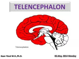
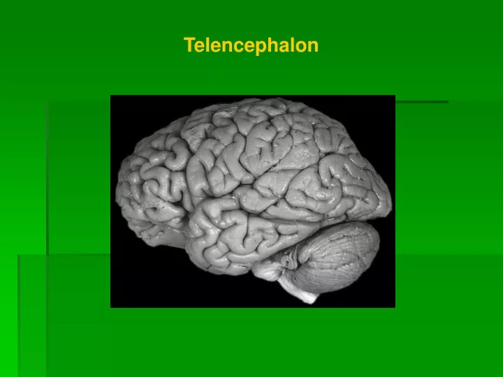
Telencephalon. Structure of telencephalon. Gray matter Cortex (pallium) Basal ganglia (striatum). White matter - pathways Projection Commissural Association. neocortex. archicortex. palleocortex. Cerebral cortex. ALLOCORTEX 3- 5 layers a) palleocortex (rhinencephalon)

E N D
Structure of telencephalon Gray matter Cortex (pallium) Basal ganglia (striatum) White matter - pathways Projection Commissural Association
neocortex archicortex palleocortex Cerebral cortex ALLOCORTEX 3-5 layers a) palleocortex(rhinencephalon) b) archicortex(limbic system) MESOCORTEX = peripaleocortex, periarchicortex NEOCORTEX (ISOCORTEX) 6 layers
Limbic lobe Rhinencephalon Bulbus olfactorius Tractus olfactorius Tuberculum olf. Stria olf. med. et lat. Gyrus cinguli Gyrus parahippocampalis Indusium griseum
ARCHICORTEX Hippocampal complex: Hippocampus (cornu ammonis, CA) Gyrus dentatus Subiculum
gyrus cinguli Limbic system – classic conception Papez‘s circuit (James Papez 1939) without specific function ncl. anterior thalami tr. mammilo-thalamicus ncl. mamillaris fornix gyrus parahippocampalis hippocampus
RECENT CONCEPTION OF LIMBIC FOREBRAIN • basomedial telencephalon, structures of diencephalon and • mesencephalon for emotion and motivation of our behavior Regular structures • g. cinguli, g. parahippocampalis, hippocampus, insular cortex, • neocortical regions of forebrain - basal frontotemporal • regions, orbital cortex • area septalis, amygdalar ncll., ventral striatum (pallidum) • ncl. anterior et medialis dorsalisthalami, habenulla • hypothalamus (ncl. mammillaris)
Limbic system – classic conception Papez‘s circuit (James Papez 1939)
Image of tooth pain Image of fear Reminiscence of music hearing
Brodman’s map (cytoarchitectonic map of cortex)■ 11 regiones ■52 areae
Functionalregions of cortex Primary motor c.(a 4), primary somatic sensory c.(a 3,1,2), primary visual c.(a 17), primary auditory c.(a 41,42) Secondary andassociation areas
SOMATOSENZORY „HOMUNCULUS“ MOTOR „HOMUNCULUS“
CORTICAL AREAS FOR SPEECH - I Broca‘s (motor) cortical area - g. front. inf. a44, 45 lesion - expressive aphasia – the lack of speech, but understanding is OK • Wernicke‘s (sensory) cortical area - a 22,39,40 in dominant hemi- • sphere • lesion - receptive aphasia – the lack of understanding
Functionalregions of cortex Secondary andassociation areas
BASAL GANGLIA AND RELATED STRUCTURES ncl. caudatus, putamen, globus pallidus (ext. + int. segment), claustrum and amygdalar ncll. Functionally: + thalamus, substantia nigra, ncl. subthalamicus, ventral tegmental area (VTA) globus pallidus + putamen = ncl. lentiformis
1 4 3 5 2 6 Basal ganglia 1 ncl. caudatus 2 globus pallidus 3putamen 4 claustrum ■corp. amygdaloideum Functionally 5ncl. subthalamicus6 substantia nigra
Corpus amygdaloideum 1) behavior for preservation of self 2) learning 3) emotion processing
Development of BG Neostriatum (striatum) ncl. caudatus, putamen – dorsal striatum ncl. accumbens – ventral striatum Palleostriatum (pallidum) = globus palliduslat. + med. segment – dorsal pallidum ventral pallidum Archistriatum corpus amygdaloideum
C Pu GP VP VS Ncl caudatus + putamen = dorsal striatum Globus pallidus = dorsal pallidum Substantia innominata: VS = ventr. striatum (ncl. accumbens septi) VP = ventral pallidum Ncl. basalis Meynerti
Functional connections of BG CORTEX STRIATUM THALAMUS PALLIDUM Function of BG inhibition of cortical and subcortical motor functions
NCL. CAUDATUS • obsessive compulsive disorder (OCD) • attention deficit disorder (ADD) • depression • schizophrenia • PAP syndrome • Huntington's disease PUTAMEN • Tourette's syndrome NCL. BASALIS • Alzheimer’s disease SUBSTANTIA NIGRA • Parkinson’s disease
centrum semiovale White matter of the telencephalon - corpus medullare Fibers commissuralprojection association
1 3 2 Commissural fibers 1corpus callosumneocortex 2commissura ant. pars ant.- paleocortex pars post. - neocortex 3commissura fornicis archicortex
Corpus callosum - 300 million fibers forceps minorconnection of frontal lobes forceps majorconnection of occipital lobes tapetum roof of the posterior horn
Projection fibers short ■ connections between cortex and BG ■reciprocal connections between cortex and thalamus long tr. co-sp tr. co-ncl tr. co-ret tr. co-tec tr. co-ru tr. co-bulb tr. co-po capsula interna
CAPSULA INTERNA crus anterius –tr. thalamo-corticalis ant. and tr. fronto-pontinus genu- tr. cortico-nuclearis(from area 4 to contralateral motoneurons of cranial nerves) crus posterius - tr. cortico-spinalis (somatotopic arrangement), tr. cortico-reticularis and tr. cortico-rubralis, tr. thalamo-corticalis posterior (somatosenzory information to parietal cortex), tr. parieto-, temporo-, occipito-pontinus,radiatio optica, radiatio acustica
crus ant. fr-po co-ncl genu co-sp, ru,re p,o,t-po crus post. radiatio acustica radiatio optica
Association fibers: short (fibrae arcuatae), long(fasciculus longitudinalis sup. et inf., fasciculus occipitofrontalis sup., fasciculi occipitales verticales, fasciculus uncinatus, cingulum) fibrae arcuatae cingulum f. longit. sup. f. uncinatus f. longit. inf.
Illustrations were copiedfrom: • Atlas der Anatomie des Menschen/ Sobotta. Putz,R., und Pabst,R. 20. Auflage. München: Urban & Schwarzenberg, 1993 • Netter: Interactive Atlas of Human Anatomy. Windows Version 2.0