Download
1 / 61
610 likes | 869 Vues
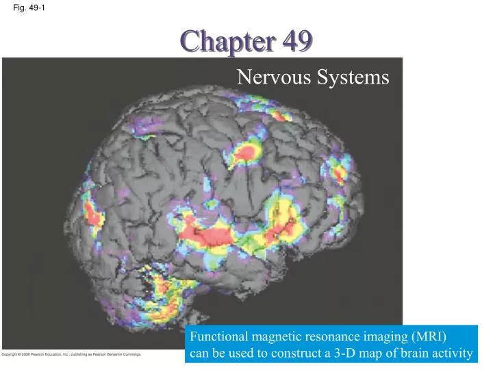
Chapter 49. Nervous Systems. Fig. 49-1. Functional magnetic resonance imaging (MRI) can be used to construct a 3-D map of brain activity. Fig. 49-2. Eyespot. Brain. Brain. Radial nerve. Nerve cords. Ventral nerve cord. Nerve ring. Transverse nerve. Nerve net. Segmental

E N D
Chapter 49 Nervous Systems Fig. 49-1 Functional magnetic resonance imaging (MRI) can be used to construct a 3-D map of brain activity
Fig. 49-2 Eyespot Brain Brain Radial nerve Nerve cords Ventral nerve cord Nerve ring Transverse nerve Nerve net Segmental ganglia (a) Hydra (cnidarian) (b) Sea star (echinoderm) (d) Leech (annelid) (c) Planarian (flatworm) Brain Brain Ganglia Anterior nerve ring Spinal cord (dorsal nerve cord) Ventral nerve cord Brain Sensory ganglia Longitudinal nerve cords Ganglia Segmental ganglia (e) Insect (arthropod) (h) Salamander (vertebrate) (f) Chiton (mollusc) (g) Squid (mollusc)
Cell body of sensory neuron in dorsal root ganglion Gray matter Quadriceps muscle a knee-jerk reflex White matter Hamstring muscle Spinal cord (cross section) Sensory neuron Motor neuron Interneuron
Peripheral nervous system (PNS) Central nervous system (CNS) Brain Cranial nerves Fig. 49-4 Spinal cord Ganglia outside CNS Spinal nerves • The CNS is composed of the brain and spinal cord • The peripheral nervous system (PNS) is composed of nerves and ganglia
Gray matter Fig. 49-5 • cerebrospinal fluid neuron cell bodies, dendrites, and unmyelinated axons White matter • bundles of myelinated axons Ventricles
PNS CNS Astrocyte Neuron VENTRICLE Oligodendrocyte Ependy- mal cell Fig. 49-6 Schwann cells Microglial cell Capillary (a) Glia in vertebrates (b) Astrocytes (LM)
◇ Epidermal cells promote circulation of cerebrospinal fluid ◇ Microglia protect the nervous system from microorganisms ◇ Oligodendrocytes and Schwann cells form the myelin sheaths around axons ◇ Astrocytes provide structural support for neurons, regulate extracellular ions and neurotransmitters, and induce the formation of a blood-brain barrier that regulates the chemical environment of the CNS ◇ Radial glia play a role in the embryonic development of the nervous system Glia have numerous functions
CNS PNS Neuron VENTRICLE Astrocyte Fig. 49-6a Ependy- mal cell Oligodendrocyte Schwann cells Microglial cell Capillary (a) Glia in vertebrates (b) Astrocytes (LM)
The Peripheral Nervous System PNS Fig. 49-7-2 Afferent (sensory) neurons Efferent neurons Autonomic nervous system Motor system Hearing carries signals to skeletal muscles and is voluntary controls activity of the digestive tract, pancreas, and gall bladder antagonistic effects Sympathetic division Parasympathetic division Enteric division Locomotion fight-or-flight” response “rest and digest Hormone action Gas exchange Circulation Digestion
Sympathetic division Parasympathetic division Action on target organs: Action on target organs: Dilates pupil of eye Constricts pupil of eye Fig. 49-8 Inhibits salivary gland secretion Stimulates salivary gland secretion Sympathetic ganglia Constricts bronchi in lungs Relaxes bronchi in lungs Cervical Slows heart Accelerates heart Stimulates activity of stomach and intestines Inhibits activity of stomach and intestines Thoracic Stimulates activity of pancreas Inhibits activity of pancreas Stimulates glucose release from liver; inhibits gallbladder Stimulates gallbladder Lumbar Stimulates adrenal medulla Promotes emptying of bladder Inhibits emptying of bladder Sacral Promotes erection of genitals Promotes ejaculation and vaginal contractions Synapse
Parasympathetic division Sympathetic division Action on target organs: Action on target organs: Fig. 49-8a Dilates pupil of eye Constricts pupil of eye Inhibits salivary gland secretion Stimulates salivary gland secretion Sympathetic ganglia Constricts bronchi in lungs Cervical Slows heart Stimulates activity of stomach and intestines Stimulates activity of pancreas Stimulates gallbladder
Sympathetic division Parasympathetic division Relaxes bronchi in lungs Fig. 49-8b Accelerates heart Inhibits activity of stomach and intestines Thoracic Inhibits activity of pancreas Stimulates glucose release from liver; inhibits gallbladder Lumbar Stimulates adrenal medulla Promotes emptying of bladder Inhibits emptying of bladder Sacral Promotes erection of genitals Promotes ejaculation and vaginal contractions Synapse
Cerebrum (includes cerebral cortex, white matter, basal nuclei) Telencephalon Fig. 49-9 Forebrain Diencephalon Diencephalon (thalamus, hypothalamus, epithalamus) Midbrain Mesencephalon Midbrain (part of brainstem) Metencephalon Pons (part of brainstem), cerebellum Hindbrain Myelencephalon Medulla oblongata (part of brainstem) Diencephalon: Cerebrum Mesencephalon Hypothalamus Metencephalon Thalamus Midbrain Pineal gland (part of epithalamus) Myelencephalon Hindbrain Diencephalon Brainstem: Midbrain Pons Spinal cord Pituitary gland Forebrain Medulla oblongata Telencephalon Spinal cord Cerebellum Central canal (c) Adult (a) Embryo at 1 month (b) Embryo at 5 weeks
Telencephalon Forebrain Diencephalon Fig. 49-9ab Mesencephalon Midbrain Metencephalon Hindbrain Myelencephalon Mesencephalon Metencephalon Midbrain Myelencephalon Hindbrain Diencephalon Spinal cord Forebrain Telencephalon (a) Embryo at 1 month (b) Embryo at 5 weeks
Cerebrum (includes cerebral cortex, white matter, basal nuclei) Diencephalon (thalamus, hypothalamus, epithalamus) Fig. 49-9c Midbrain (part of brainstem) Pons (part of brainstem), cerebellum Medulla oblongata (part of brainstem) Diencephalon: Cerebrum Hypothalamus Thalamus Pineal gland (part of epithalamus) Brainstem: Midbrain Pons Pituitary gland Medulla oblongata Spinal cord Cerebellum Central canal (c) Adult
Fig. 49-UN1 • The brainstem has three parts: the midbrain, the pons, and the medulla oblongata
The midbrain contains centersfor receipt and integration of sensory information The pons regulates breathing centersin the medulla The medulla oblongata contains centers that control several functions including breathing, cardiovascular activity, swallowing, vomiting, and digestion
The brainstem and cerebrum control arousal and sleep The core of the brainstem has a diffuse network of neurons called the reticular formation This regulates the amount and type of information that reaches the cerebral cortex and affects alertness The hormonemelatonin is released by the pineal gland and plays a role in bird and mammal sleep cycles Arousal and Sleep-reticular formation
Fig. 49-10 Eye Input from nerves of ears Reticular formation Input from touch, pain, and temperature receptors
Sleep is essential and may play a role in the consolidation of learning and memory Dolphins sleep with one brain hemisphere at a time and are therefore able to swim while “asleep” Fig. 49-11 Low-frequency waves characteristic of sleep High-frequency waves characteristic of wakefulness Time: 1 hour Location Time: 0 hours Left hemisphere Right hemisphere
The cerebellum ---is important for coordination and error checking during motor, perceptual, and cognitive functions ---It is also involved in learning and remembering motor skills Fig. 49-UN2
Diencephalon The diencephalon develops into three regions: the epithalamus, thalamus, and hypothalamus ---The epithalamus includes the pineal gland and generates cerebrospinal fluid from blood ---The thalamus is the main input center for sensory information to the cerebrum and the main output center for motor information leaving the cerebrum ---The hypothalamus regulates homeostasis and basic survival behaviors such as feeding, fighting, fleeing, and reproducing Fig. 49-UN3
The hypothalamus also regulates circadian rhythms such as the sleep/wake cycle Mammals usually have a pair of suprachiasmatic nuclei (SCN) in the hypothalamus that function as a biological clock Biological clocks usually require external cues to remain synchronized with environmental cycles Biological Clock Regulation by the Hypothalamus
RESULTS Wild-type hamster hamster Wild-type hamster with SCN from hamster hamster with SCN from wild-type hamster Fig. 49-12 24 23 22 Circadian cycle period (hours) 21 20 19 After surgery and transplant Before procedures
Cerebrum The cerebrum has right and left cerebral hemispheres ---Each cerebral hemisphere consists of a cerebral cortex (gray matter) overlying white matter and basal nuclei ---In humans, the cerebral cortex is the largest and most complex part of the brain ---The basal nuclei are important centers for planning and learning movement sequences ---A thick band of axons called the corpus callosumprovides communication between the right and left cerebral cortices ---The right half of the cerebral cortex controls the left side of the body, and vice versa • The cerebrum develops from the embryonic telencephalon
Fig. 49-13 Right cerebral hemisphere Left cerebral hemisphere Thalamus Corpus callosum Basal nuclei Cerebral cortex
The outermost layer of the cerebral cortex has a different arrangement in birds and mammals In mammals, the cerebral cortex has a convoluted surface called the neocortex,which was previously thought to be required for cognition Cognition is the perception and reasoning that form knowledge However, it has recently been shown that birds also demonstrate cognition even though they lack a neocortex Evolution of Cognition in Vertebrates
Fig. 49-14 Pallium Cerebral cortex Cerebrum Cerebrum Cerebellum Cerebellum Thalamus Thalamus Midbrain Midbrain Hindbrain Hindbrain Avian brain to scale Human brain Avian brain
Each side of the cerebral cortex has four lobes: frontal, temporal, occipital, and parietal Each lobe contains primary sensory areas and association areas where information is integrated Thalamus directs different types of inputs to distinct locations Primary sensory areas association areasthe frontal association area (plan actions and movement) motor cortex (at the rear of the frontal lobe) brain stem Spinal cord Concept 49.3: The cerebral cortex controls voluntary movement and cognitive functions
Thalamus directs different types of inputs to distinct locations Frontal lobe: olfactory information Parietal lobe: somatosensory input, tastes Fig. 49-15 Somatosensory cortex Motor cortex Somatosensory association area Speech Frontal association area Taste Reading Speech Hearing Visual association area Smell Auditory association area Vision Primary sensory areas association areasthe frontal association area (plan actions and movement) motor cortex (at the rear of the frontal lobe) brain stem Spinal cord Temporal lobe: auditory Occipital lobe: visual
The cerebral cortex receives input from sensory organs and somatosensory receptors (touch, pain, pressure, temperature, and the position of muscles and limbs) Specific types of sensory input enter the primary sensory areas of the brain lobes Adjacent areas process particular features in the sensory input and integrate information from different sensory areas In the somatosensory and motor cortex, neurons are distributed according to the body part that generates sensory input or receives motor input Information Processing in the Cerebral Cortex
Parietal lobe Frontal lobe Fig. 49-16 Upper arm Shoulder Trunk Head Knee Leg Trunk Neck Hip Elbow Hip Forearm Elbow Wrist Forearm Hand Hand Fingers Fingers Thumb Thumb Eye Neck Nose Brow Face Eye Lips Genitals Toes Face Teeth Gums Jaw Lips Jaw Tongue Tongue Pharynx Primary motor cortex Primary somatosensory cortex Abdominal organs Neurons are distributed in an orderly fashion
Shoulder Knee Trunk Elbow Forearm Hip Wrist Hand Fig. 49-16a Fingers Thumb Neck Brow Eye Toes Face Lips Jaw Tongue Primary motor cortex
Upper arm Trunk Neck Leg Head Hip Elbow Forearm Fig. 49-16b Hand Fingers Thumb Eye Nose Face Lips Genitals Teeth Gums Jaw Tongue Pharynx Primary somatosensory cortex Abdominal organs
1.失去說話的能力稱做「失語症」。古希臘人發現,腦部損傷會導致失語症。 幾世紀以後,1836 年Marc Dax 描述了一組不能適當地講話的的病患。 Dax 報道所有這些患者他們的腦子的左邊都有損傷。1861 年又過了四分之一世紀以後,保羅˙布洛卡(Paul Broca )描述了只能說一個字‘’Tan‘’的一名患者 2. 1876年 卡爾˙韋尼克(Karl Wernicke)發現對腦子的另一個區域的損傷也會造成語言問題。 「韋尼克氏區」(Wernicke's area)是在布洛卡區比較後方集下方的位置。 實際上, 韋尼克氏區是在顳葉後方的位置。 布洛卡區和韋尼克氏區由神經纖維束「arcuate fasciculus 」連接稱。 arcuate fasciculus 損傷導致的病症叫做「傳導失語症」。
Max language and speech Wernicke’s area: understsnding language but unable to speak (left temporal lobe) Fig. 49-17 Reading w/o speaking Hearing words Seeing words Generates verbs Min Generating words Speaking words Broca’s area: left frontal lobe comprehend speech but not the ability to speak
The corpus callosum transmits information between the two cerebral hemispheres The left hemisphere is more adept at language, math, logic, and processing of serial sequences The right hemisphere is stronger at pattern recognition, nonverbal thinking, and emotional processing The differences in hemisphere function are called lateralization Lateralization is linked to handedness Lateralization of Cortical Function
Emotions are generated and experienced by the limbic system and other parts of the brain including the sensory areas The limbic system is a ring of structures around the brainstem that includes the amygdala, hippocampus, and parts of the thalamus The amygdala is located in the temporal lobe and helps store an emotional experience as an emotional memory Emotions
Thalamus Hypothalamus Fig. 49-18 Prefrontal cortex Olfactory bulb Hippocampus Amygdala
Two processes dominate embryonic development of the nervous system Neurons compete for growth-supporting factors in order to survive Only half the synapses that form during embryo development survive into adulthood` Neural plasticity describes the ability of the nervous system to be modified after birth Changes can strengthen or weaken signaling at a synapse Concept 49.4 Changes in synaptic connections underlie memory and learning
N1 N1 Fig. 49-19 N2 N2 (a) Synapses are strengthened or weakened in response to activity. (b) If two synapses are often active at the same time, the strength of the postsynaptic response may increase at both synapses.
Learning can occur when neurons make new connections or when the strength of existing neural connections changes Short-term memory is accessed via the hippocampus The hippocampus also plays a role in forming long-term memory, which is stored in the cerebral cortex Memory and Learning
In the vertebrate brain, a form of learning called long-term potentiation (LTP)長期增益現象 involves an increase in the strength of synaptic transmission LTP involves glutamate receptors If the presynaptic and postsynaptic neurons are stimulated at the same time, the set of receptors present on the postsynaptic membranes changes Long-Term Potentiation
Fig. 49-20 Ca2+ Long-term potentiation in the brain Na+ Mg2+ blocked NMDA glutamate receptor Mg2+ Glutamate NMDA receptor (closed) AMPA (α-amino-3-hydroxyl-5-methyl-4-isoxazole-propionate) NMDA receptor (open) Stored AMPA receptor • Depolarizes Mg2+ removed • Respond to glutamate an influx of Na+ and Ca2+ • Ca2+AMPA glutamate receptors inserted into the postsynaptic membrane (a) Synapse prior to long-term potentiation (LTP) 1 3 2 (b) Establishing LTP • Glutamate release Na+ influx depolarization • Depolarization • Mg2+ removed • AMPA and NMPA receptor trigger postsynaptic potentials 3 4 1 2 (c) Synapse exhibiting LTP
AMPA AMPA (α-amino-3-hydroxyl-5-methyl-4-isoxazole-propionate) is a compound that is a specific agonist for the AMPA receptor, where it mimics the effects of the neurotransmitterglutamate.[ NMDA (N-methyl-D-aspartic acid) is an amino acid derivative acting as a specific agonist at the NMDA receptor, and therefore mimics the action of the neurotransmitterglutamate on that receptor. In contrast to glutamate, NMDA binds to and regulates the above receptor only, but not other glutamate receptors
Disorders of the nervous system include schizophrenia, depression, Alzheimer’s disease, and Parkinson’s disease Genetic and environmental factors contribute to diseases of the nervous system Concept 49.5: Nervous system disorders can be explained in molecular terms
About 1% of the world’s population suffers from schizophrenia Schizophrenia is characterized by hallucinations, delusions 錯覺, blunted emotions, and other symptoms Available treatments focus on brain pathways that use dopamine as a neurotransmitter Schizophrenia
50 Genes shared with relatives of person with schizophrenia 12.5% (3rd-degree relative) Fig. 49-21 40 25% (2nd-degree relative) 50% (1st-degree relative) 100% 30 Risk of developing schizophrenia (%) 20 10 0 Parent Child Uncle/aunt Grandchild First cousin Half sibling Full sibling Identical twin Nephew/niece Fraternal twin Individual, general population Relationship to person with schizophrenia
Two broad forms of depressive illness are known: major depressive disorder and bipolar disorder In major depressive disorder, patients have a persistent lack of interest or pleasure in most activities Bipolar disorder is characterized by manic (high-mood) and depressive (low-mood) phases Treatments for these types of depression include drugs such as Prozac and lithium Depression