Download

1 / 102
1.09k likes | 1.39k Vues
Role of Interventional Radiology in a Gastrointestinal Tumor Service. Anastasia Balius, MD Interventional Radiology UTMCK October 23, 2011. Hepatocellular Carcinoma. Approximately 24,120 persons diagnosed with liver or intrahepatic bile duct cancer in the US in 2010 1

E N D
Role of Interventional Radiology in a Gastrointestinal Tumor Service Anastasia Balius, MD Interventional Radiology UTMCK October 23, 2011
Hepatocellular Carcinoma • Approximately 24,120 persons diagnosed with liver or intrahepatic bile duct cancer in the US in 20101 • 18,910 deaths from those cancers during that year1 • Incidence has risen significantly in developed countries in the past two decades2 • Increased prevalence of hepatitis B and C 1American Cancer Society. Statistics last revised 7/7/10 2Ince N, Wands JR. The increasing incidence of heaptocellular carcinoma. N Engl J Med 1999;340:798-799. .
Risk Factors • Same as those for Cirrhosis • Hepatitis B/C • Inherited errors of metabolism • Autoimmune hepatits • Non -alcoholic steatohepatitis [NASH] • Excessive alcohol intake • Environmental exposure to aflatoxin 3
Hepatocellular Carcinoma (HCC) • With definitive treatment 5 year survival is over 50%1 • For all stages combined, 5 year survival is approximately 10%1 • Only 10-15% of patients are candidates for curative therapy2,3 • Surgical resection or transplant 1American Cancer Society. Statistics last revised 8/16/10 2Llovet JM. Treatment of hepatocellular carcinoma. Curr Treat Options Gastroenterol. 2004;7:431-441 3Kanematsu T, Furui J, Yanaga K, et al. A 16-year experience in performing hepatic resection in 303 patients with hepatocellular carcinoma. J Vasc Interv Radiol 1995; 6:71–74.
Metastatic Colorectal Cancer (mCRC) • 5 year survival for stage IV colorectal cancer is 6%1 • Liver is most frequent site of metastases • Approximately 60% of patients with mCRC will eventually have liver as predominant site of disease2 • Surgical resection is treatment of choice • 5 year survival rates after resection >50%3,4 • Feasible in <20% of patients2 1American Cancer Society. Statistics last revised 3/2/2011 2Sasson AR, Sigurdson ER. Surgical treatment of liver metastases. Semin Oncol. 2002;29:107-118 3Choti MA, et al. Trends in long-term survival following liver resection for hepatic colorectal metastases. Ann Surg. 2002;235:759-766. 4Pawlik TM, et al. Effect of surgical margin status on survival and site of recurrence after hepatic resection for colorectal metastases. Ann Surg. 2005;241:715-722, discussion 722-714.
NCCN Categories of evidence • Category 1: The recommendation is based on high-level evidence (eg randomized controlled trials) and there is uniform NCCN consensus • Category 2A: The recommendation is based on lower-level evidence and there is uniform NCCN consensus • Category 2B: The recommendation is based on lower-level evidence and there is non uniform NCCN consensus (but no major disagreement) • Category 3: The recommendation is based on any level of evidence but reflects major disagreement • All recommendations are Category 2A unless otherwise noted
Treatment of HCC Potentially resectable or transplantable, operable by performace status or comorbidity HCC confirmed • Multidisciplinary Evaluation • H+P • Hepatitis Panel • Bilirubin, transaminases, alk phos, LDH • PT /INR, albumin, protein, BUN, Cr • CBC, platelets • AFP • Chest imaging • Bone scan as indicated Metastatic disease Inoperable by performance status or comorbidity, local disease only Unresectable
Treatment of HCC For patients with an estimated FLR/total liver volume ratio below recommended values who are otherwise suitable candidates for liver resection, pre-operative portal vein embolization (PVE) should be considered NCCN Practice Guidelines in Oncology – v.2.2011, Hepatobiliary Cancers.
Treatment of HCC Clinical presentation Surgical Assessment Treatment • Child’s A, B • No portal hypertension • Suitable tumor location • Adequate liver reserve • Suitable liver remnant Resection, if feasible (preferred) or locoregional therapy Potentially resectable or transplantable • UNOS criteria • Patient has a tumor <5 cm in dm or 2-3 tumors < 3 cm each • No macrovascular involvement • No extrahepatic disease • These patients may be resected if transplantation not feasible Liver Transplant
Child-Pugh Score Clinical/Biochemical Parameters Points for Increasing Abnormality Class A = 5-6 points; Class B = 7 -9 points; Class C = 10-15 points
Treatment of HCC Clinical Presentation Treatment Options: Sorafenib (Child-Pugh Class A [Category 1] or B) Clinical Trial Locoregional Therapy RT (conformal or sterotactic) (category 2B) Supportive Care Inoperable by performance status or comorbity, local disease only Sorafenib (Child-Pugh Class A [Category 1] or B) Or Supportive Care Or Clinical trial Metastatic disease
Locoregional Therapy Ablation Embolization • Tumor and margin of normal tissue should be treated • Tumors < 3 cm; lesions between 3-5 cm should be treated in combination with embolization • Arterial blood supply to the tumor must be able to be isolated without non-target embolization • Relatively contraindicated with bilirubin >3 mg/dL • Contraindicated with main PV thrombosis (relative) or Child-Pugh Class C (absolute)
Treatment of HCC • Inadequate hepatic reserve • Tumor location Transplant Meets UNOS criteria? Not a transplant candidate Unresectable Options: Sorafenib (Child-Pugh Class A [Category 1] or B) Chemotherapy + RT only in the context of a clinical trial Clinical trial Locoregional therapy RT (conformal or sterotactic) (category 2B) Supportive care Systemic or intra-arterial chemotherapy in clinical trial Extensive liver tumor burden
Treatment of mCRC When hepatic disease is not optimally resectable based on insufficient remnant liver volume, approaches utilizing preoperative portal vein embolization1 or staged liver resection can be considered NCCN Practice Guidelines in Oncology – v.3.2011, Colon Cancer 1Covey AM, et al. Combined portal vein embolization and neoadjuvant chemotherapy as a treatment strategy for resectable hepatic colorectal metastases. Ann Surg. 2008 Mar;247(3):451-5.
Treatment of mCRC • Ablative techniques may be considered alone or in conjuction with resection. All original sites of disease need to be amenable to ablation or resection. • Some institutions use arterially directed embolic therapy in highly select patients with chemotherapy resistant/refractory disease without obvious systemic disease, with predominent hepatic metastases (category 3)
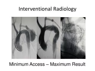
Interventional Radiology’sRole • Supportive procedures for the oncology patient • Determination of disease • Facilitation of definitive surgical treatment • Treatment of non-surgical candidates
Determination of Disease • AFP and ultrasound are used as screening for HCC • Additional imaging is indicated in the setting of a rising serum AFP or identification of a liver mass nodule on US
Determination of Disease • Three different modalities • Triphasic helical CT • Triphasic dynamic contrast enhanced MRI • Contrast enhanced U/S
Determination of Disease • Nodules 1 to 2 cm in size need to demonstrate classic arterial enhancement with two different diagnostic techniques • Nodules > 2 cm need demonstrate classic arterial enhancement with only one diagnostic technique • Nodules < 1 cm should be re-evaluated every 3 to 4 months until fit into a size criteria • If nodules are non-diagnostic on imaging, tissue sampling is needed
Facilitation of Definitive Surgical Treatment Portal Vein Embolization Transplant “Bridge”
Portal Vein Embolization (PVE) • In primary or secondary hepatic malignancy, extended hepatectomies provide a chance of cure1 • Morbidity and mortality of such procedures are considerable2 • Volume and function of the liver remnant is a major risk factor for perioperative complications3,4 1Vauthey JN, et al. Is extended hepatectomy for hepatobiliary malignancy justified? Annals of Surgery. 2004 May; 239(5):722-30; discussion 30-2 2Jarnagin WR, et al. Improvement in perioperative outcome after hepatic resection: analysis of 1,803 consecutive cases over the past decade. Annals of Surgery. 2002 Oct;236(4):397-406; discussion-7.. 3Abdalla EK, et al. Extended hepatectomy in patients with hepatobiliary malignancies with and without preoperative portal vein embolization. Arch Surg. 2002 Jun:137(6):675-80; discussion 80-1 4Vauthey JN, et al. Standardized measurement of the future liver remnant prior to extended liver resection: methodology and clinical associations. Surgery. 2000 May;127(5):512-9
Portal Vein Embolization • PVE induces hypertrophy in the non-embolized liver segments • Increase in volume and function of future liver remnant (FLR) • Decreases risk of post-operative hepatic insufficiency Abdalla EK, et al. Portal vein embolization: rationale, technique and future prospects. The British Journal of Surgery. 2001 Feb;88(2):165-75 Makuuchi M, et al. Preoperative portal embolization to increase safety of major hepatectomy for hilar bile duct carcinoma: a preliminary report. Surgery. 1990 May;107(5):521-7
PVE • Normal underlying liver1 • FLR should be 20-25% of total liver volume (TLV) • Chemotherapy induced liver injury2 • FLR should be >30% of TLV • Chronic liver disease (cirrhosis or severe fibrosis)2 • FLR should be >40% 1Abdalla EK, et al. Extended hepatectomy in patients with hepatobiliary malignancies with and without preoperative portal vein embolization. Arch Surg. 2002 Jun;137(6):675-80; discussion 80-1. 2Azoulay D, et al. Percutaneous portal vein embolization increases the feasibility and safety of major liver resection for hepatocellular carcinoma in injured liver. Annals of Surgery. 2000 Nov;232(5):1176-81.
PVE Contraindications • Extrahepatic metastases • Overt portal hypertension • Tumor invasion of the portal vein • Tumor invasion of the FLR • Biliary obstruction • Renal insufficiency • Uncorrectable coagulopathy
PVE • Performed under conscious sedation • 2 standard approaches • Percutaneous ipsilateral • Percutaneous contralateral • Wide array of embolic agents • Coils, amplatzer plugs • Particles • Absolute alcohol • Fibrin glue • N-BCA (n-butyl cyanoacrylate)
Complications • Bleeding (0.2 – 1/6%) • Pneumothorax • Sepsis • Liver abscess (0.3%) • Portal hypertension resulting in esophageal variceal hemorrhage • Migration of embolic material (0.2-5.3%) • Portal vein thrombosis of FLR (<1%) • Portal vein dissection • Intraparenchymal injury • Transient liver failure (3.2%) Di Stefano DR, et al. Preoperative percutaneous portal vein embolization: evaluation of adverse events in 188 patients. Radiology. 1993 Jul;188(1):73-7 Kodama Y, et al. Complications of percutaneous transhepatic portal vein embolization. J Vasc Interv Radiol. 2002 Dec;13(12):1233-7
Outcomes • Average expected growth of FLR = 8-27%1 • 75% of growth occurs in first 3 weeks post-PVE2 • Failure to hypertrophy >5% following PVE is associated with a significantly higher risk of major complications, hepatic insufficiency, and increased 90-day mortality2 1Abulkhir A, et al. Preoperative portal vein embolization for major liver resection: a meta-analysis. Ann Surg. 2008 Jan;247(1):49-57 2Ribero D, et al. Portal vein embolization before major hepatectomy and its effects on regeneration, resectability, and outcome. The British Journal of Surgery. 2007 Nov;94(11): 1386-94
Facilitation of Definitive Surgical Treatment • Locoregional treatment of HCC as a “bridge” to liver transplantation1,2 • Radiofrequency ablation3,4 • Chemoembolization5 • Radioembolization 1Bruix J, Sherman M. Management of hepatocellular carcinoma. Hepatology. 2005;42:1208-1236 2Llover JM, et al. Design and endpoints of clinical trials in hepatocellular carcinoma. J Natl Cancer Inst. 2008;100:698-711. 3Pompili M, et al. Percutaneous ablation procedures in cirrhotic patients with hepatocellular carcinoma submitted for liver transplantation: assessment of efficacy at explant analysis and of safety for tumor recurrence. Liver Transpl. 2005;11:1117-1126. 4Mazzaferro V, et al. Radiofrequency ablation of small hepatocellular carcinoma in cirrhotic patients awaiting liver transplantation: a prospective study. Ann Surg. 2004;240:900-909. 5Richard HM, 3rd, et al. Hepatic arterial complications in liver transplant recipients treated with pretransplantation chemoembolization for hepatocellular carcinoma. Radiology. 2000;214:775-779.
Treatment of Nonsurgical Candidates • Ablative techniques • Radiofrequency ablation • Microwave ablation • Alcohol ablation • Arterial embolization • Trans-arterial embolization (TAE) • Trans-arterial chemoembolization (TACE) • Trans-arterial radioembolization or Selective Internal Radiation Therapy (SIRT) • Combination therapy
Patient Work-up • Laboratory studies • CBC, Chem 7, PT/PTT, liver function tests, tumor markers • Cross-sectional imaging (CT/MRI) • Childs class • Performance status • ECOG/Karnofsky • Staging • Consent with emphasis on palliation and management of expectations
Alcohol versus RFA • RFA was shown to be superior to PEI with respect to complete response rate1 • RFA demostrated a lower rate of recurrence2,3 • Overall survival higher for RFA2,3 • Fewer treatment sessions for RFA3 1Brunello F, et al. Radiofrequency ablation versus ethanol injection for early hepatocellular carcinoma: A randomized controlled trial. Scand J Gastroenterol. 2008;43:727-735 2Lin SM, et al. Radiofrequency ablation improves prognosis compared with ethanol injection for hepatocellular carcinoma <4 cm. Gastroenterology. 2004;127:1714-1723 3Shiina S, et al. A randomized controlled trial of radiofrequency ablation with ethanol injection for small hepatocellular carcinoma. Gastroenterology. 2005;129:122-130.
RFA vs. Ethanol Injection Survival rate 1Lencioni R, et al. Small hepatocellular carcinoma in cirrhosis: randomized comparison of radiofrequency ablation versus percutaneous ethanol injection. Radiology 2003;228:235-240. 2Lin SM, et al. Radiofrequency ablation improves prognosis compared with ethanol injection for hepatocellular carcinoma < or = 4 cm. Gastroenterology 2004;127:1714-1723. 3Shiina S, et al. A randomized controlled trial of radiofrequency ablation versus ethanol injection for small hepatocellular carcinoma. Gastroenterology. 2005;129:122-130.
Local recurrence rates for HCC • More recent study found <3% of patients with single HCC tumor < 2 cm had recurrent disease at 31 months after repeated application of RFA1 1Livraghi T, et al. Sustained complete response and complications rates after radiofrequency ablation of very early hepatocellular carcinoma in cirrhosis: Is resection still the treatment of choice? Hepatology. 2008;47:82-89. 18
Long-Term Survival Survival Rate (%) Lencioni R, et al. Early-stage hepatocellular carcinoma in cirrhosis: long-term results of percutaneous image-guided radiofrequency ablation. Radiology 2005;234:961-967 Tateishi R, et al. Percutaneous radiofrequency ablation for hepatocellular carcinoma. Cancer 2005;103:1201-1209 Cabassa P, et al. Radiofrequency ablation of hepatocellular carcinoma: long-term experience with expandable needle electrodes. AJR 2006;185:S316-321 Choi D, et al. Percutaneous radiofrequency ablation for early-stage hepatocellular carcinoma as a first-line treatment: long-term results and prognostic factors in a large singe-institution series. Eur Radiol 2007;17:684-692.
Radiofrequency Ablation vs. Resection • RFA compared to liver resection in a prospective randomized controlled study • Patients with solitary HCC <5 cm in dm • No differences in recurrence-free survival or overall survival were found when treatment arms were compared. Chen MS, et al. A prospective randomized trial comparing percutaneous local ablative therapy and partial hepatectomy for small hepatocellular carcinoma. Ann Surg. 2006;243:321-328.
Microwave Ablation • In moderately or poorly differentiated HCC, overall survival was significantly better than with PEI1 • In study of 234 pts, 3 yr and 5 yr survival rates were 73% and 57%2 • In the one randomized trial comparing microwave and radiofrequency ablation, no statistically significant differences were observed in the efficacy of the two techniques3 1Seki T, et al. Percutaneous microwave coagulation therapy for patients with small hepatocellular carcinoma: comparison with percutaneous ethanol therapy. Cancer 1999;85:1694-1702 2Dong B, et al. Percutaneous sonographically guided microwave coagulation therapy for hepatocellular carcinoma: results in 234 patients. AJR 2003;180:1547-1541. 3Shibata T, et al. Small hepatocellular carcinoma: comparison of radio-frequency ablation and percutaneous microwave coagulation therapy. Radiology 2002;223:331-337.
RFA for mCRC 1Solbiati L, et al. Percutaneous radio-frequency ablation of hepatic metastases from colorectal cancer: long-term results in 117 patients. Radiology 2001 Oct;221(1):159-66. 2Gillams AR, Lees WR. Radio-frequency ablation of colorectal metastases in 167 patients. Eur Radiol 2004;14:2261-2267. 3Jakobs TF, et al. Radiofrequency ablation of colorectal liver metastases: mid-term results in 68 patients. Anti-cancer Res 2006;26:671-680. 4Lencioni R, et al. Percutaneous radiofrequency ablation of hepatic colorectal metastases: technique, indications, results and new promises. Invest Radiol 2004;39:689-697. 5Sorensen SM, et al. Radiofrequency ablation of colorectal liver metastases: long-term survival. Acta Radiol 2007;48:253-258.
RFA versus resection of mCRC • A number of retrospective studies have compared RFA and liver resection in the tx of liver mets1-3, although RFA has not been well studied in this setting • In retrospective studies comparing RFA and liver resection in liver mets RFA has been inferior to resection with respect to rates of local recurrence and 5-year overall survival4 • Patient selection bias, technological limitations of RFA or both?2 1Hur H, et al. Comparative study of resection and radiofrequency ablation in the treatment of solitary colorectal liver metastases. Amer J Surg. 2009;197:728-736. 2Gleisner AL, et al. Colorectal liver metastases: recurrence and survival following hepatic resection, radiofrequency ablation, and combined resection-radiofrequency ablation. Arch Surg. 2008;143:1204-1212. 3Reuter NP, et al. Radiofrequency ablation vs. resection for hepatic colorectal metastasis: therapeutically equivalent? J Gastrointest Surg. 2009;13:486-91. 4Abdalla EK. Commentary: Radiofrequency ablation for colorectal liver metastases: do not blame the biology when it is the technology. Amer J Surg. 2009;197:737-739.
Radiofrequency Ablation • Electrode probes deliver an alternating high-frequency electrical current (460 to 500 kHz) • Ion agitation is converted by friction into heat • Tissue temperature is increased • Cellular death occurs via thermal coagulation necrosis
Technique • Re-identify lesion under CT or US • Choose appropriate access site • Plan approach • Place probes • 12 to 16 minute ablation • Tract ablation
Considerations • “Heat sink” effects • Compromised sphincter of Oddi • Levaquin and flagyl prep • Adjacent structures • Diaphragm • Abdominal wall • Capsule • Bowel • Size
Development of Ablated Region • After 24 to 48 hours, necrotic lesion forms reaching maximum size by 7 days • Ablated lesion may exhibit an increase in size up to 3 months post ablation1 • Ablated tissue will be replaced by scar or reabsorbed • Treated area will not enhance on follow up imaging2 • Most residual viable tumor is evident at 1-3 months after ablation3 1McDougal WS, et al; J Urol 2005; 174:61-63 2Kawamoto, et al; Radiographics 2007; 27:343-355 3Gervais, et al; AJR 2005; 185:64-71
Complications • Hemorrhage1 • Hemoperitoneum • Hemothorax • Hemobilia • Pneumo/Hydrothorax • Abscess • Sepsis • Liver Failure2 • Tumor seeding • Skin Burn • Damage to surrounding structures • Pain in a dermatomal/ diaphragmatic distribution • Post ablation syndrome 1Goto E et al. J Clin Gastroenterol 2009 Oct 3 [Epub ahead of print} 2Kong WT, et al. World J Gastroenterol 2009 Jun 7; 15(21):2651-6 11
Complication Rate • Mortality rate ranges from 0.1% to 0.5% • Most common causes of death were sepsis and hepatic failure • Major complication rate ranges from 2.2% to 3.1% • Minor complication rate ranges from 5% to 8.9% • Most common complications were intraperitoneal bleeding, hepatic abscess, bile duct injury, hepatic decompensation, and grounding pad burns Rhim H. Complications of radiofrequency ablation in hepatocellular carcinoma. Abdom Imaging 2005 Jul-Aug; 30 (4): 409-18 Livraghi T, et al. Treatment of focal liver tumors with percutaneous radiofrequency ablation: complications encountered in a multicenter study. Radiology 2003;26:441-451. De Baere T, et al. Adverse events during radiofrequency treatment of 582 hepatic tumors. AJR 2003;181:695-700. Bleicher RJ, et al. Radiofrequency ablation in 447 complex unresectable liver tumors: lessions learned. Ann Surg Oncol 2003;10:52-58. 12