Download
1 / 29
310 likes | 360 Vues
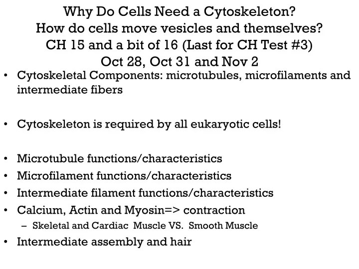
Why Do Cells Need a Cytoskeleton? How do cells move vesicles and themselves? CH 15 and a bit of 16 (Last for CH Test #3) Oct 28, Oct 31 and Nov 2. Cytoskeletal Components: microtubules, microfilaments and intermediate fibers Cytoskeleton is required by all eukaryotic cells!

E N D
Why Do Cells Need a Cytoskeleton?How do cells move vesicles and themselves?CH 15 and a bit of 16 (Last for CH Test #3)Oct 28, Oct 31 and Nov 2 • Cytoskeletal Components: microtubules, microfilaments and intermediate fibers • Cytoskeleton is required by all eukaryotic cells! • Microtubule functions/characteristics • Microfilament functions/characteristics • Intermediate filament functions/characteristics • Calcium, Actin and Myosin=> contraction • Skeletal and Cardiac Muscle VS. Smooth Muscle • Intermediate assembly and hair
Practice Quiz: Signal Transduction 1) With respect to activation of adenylyl cyclase, which G-protein subunit can be present in stimulatory or inhibitory forms? Alpha Beta Gamma Delta Omega How does a hormone receptor change the function of this subunit? 2) Which 2nd messenger is produced by phospholipase-C for the purpose of opening specific ligand-gated Ca++ channels? Diacylglycerol Calmodulin Inositol triphosphate Cyclic AMP Cyclic GMP Ca++ How is PL-C activated in this regard? 3) Which of the following is a voltage gated channel that you would expect to find at a pre-synaptic (nerve ending) where vesicles release acetylcholine (neurotransmitter). K+ Na+ Ca++ Cl- How do vesicles get from the nucleus to the end of the axon? 4) What second messenger activates protein kinase A? Diacylglycerol Calmodulin Inositol triphosphate Cyclic AMP Cyclic GMP Ca++ What enzyme creates this second messenger and what G-protein subunit is important in this regard? 5) Why do some hormones need amplification? What hormones don’t need amplification? Why don’t some hormones need second messengers? 6) How does a tyrosine kinase work? Name three examples of tyrosine kinases and their functions.
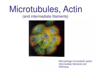
Cells must respond to stresses and changes in shape, they must also perform intracellular mechanical work. Work Requires Structure for “Leverage”: • Exocytosis/Endocytosis/Vesicle Transport • Shape changes and morphogenesis • Nuclear and cellular division The Cytoplasm has 3 types of filament: • 1-Microtubules: 25nm diameter-Largest subunit type • Movement in the cell • Tubulin: alpha/beta subunits • 2-Microfilaments: 8 nm diameter- Smallest subunit type • Movement of the cell • G-actin subunits • 3-Intermediate Filaments: 8-12 nm dia-Intermediate size • Structural support in/around cell • Tissue specific proteins These 3 filaments function to create an intracellular matrix for organization, attachment and intracellular motion.
Microtubules consist of tubulin protein dimers (α and B) that guide intracellular transport vesicles and proteins to the proper location within a cell. Sites of Special Importance: • 1-Formation of Mitotic Spindle • One Kinetochore for each chromosome • Chromosomes microtubule elongation separates the chromosomes at anaphase • 2- Guiding Vesicles: exocytosis/endocytosis • Clathrin is important especially in endocytosis • 3-Critical for axoplasmic flow in axons/dendrites • Carry vesicles with NT to axon or recycle NT to nucleus • 4- Important to structure of flagella and cilia -9+2 pattern of microtubules • 5- Inhibiting tubule synthesis kills cell (antimitotic)! • Cancer Drugs: Vinblastine: no MT formed • Taxol: MT not disassembled for division
How are microtubules made at specific nucleation cites? Alpha/Beta monomers DimersTubes(25nm dia) Tubulin-GTP is added at the positive end of the tube and removed from the negative end of tube. XXX XXX
What events inside a cell are dependent upon proper the formation/destruction of microtubules (MTs) at locations called microtubule organizing centers (MTOCs)?
Dynein and Kinesin proteins crawl along microtubules and carry vesicles with them using ATP as the energy source.Dynein: moves toward Negative Direction.Kinesin: moves toward Positive DirectionSimilar proteins form the radial spokes of flagella and cilia.
Dynein and kinesin proteins can be active at the same time and move vesicles in opposite directions (- or +) on the same microtubules!
Flagella and Cilia: Dynein arms move across adjacent MTs causing sliding/axoneme bending motion
Visualization of Dynein/ATP-dependent sliding action across adjacent axonemes in a flagella. In terms of the ATP that drive this process, why do sperm carry mitochondria? Where do your mitochondria come from? How could a genetic defect in dynein result in linkage of male sterility and respiratory disease?
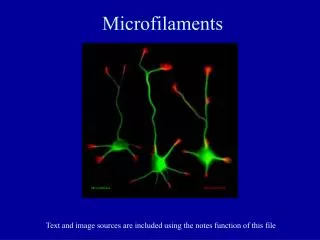
Microfilaments are the smallest cytoskeletal structures in the cell and form thread-like actin structures in the cytosol. Important for muscle contraction (actin/myosin) Important for ameboid motion/cytoplasmic streaming Organization: • Monomers of G-actin-ATP polymerize into long strands of F-actin (mature actin) • Small: about 7 nm in diameter • Strands form a double helix • Some strands become cross-linked by “filamin” for extra strength!
Microfilaments are primarily formed by adding G-actin to the positive “fast-growing” end of the filament.Actin polymerization requires: ATP, K+ and Mg++What effect does the drug cytochalasin have?
Microfilaments can form diffuse gel-like matrices OR they can become tightly cross-linked to form rigid structures like the non-motile microvilli found on intestinal epithelial cells.
Classic Link Protein Types: Ankyrin Spectrin Band 4.1 Microfilaments can also be linked to proteins in the plasma membrane and molecules/materials outside the cell itself by N-CAMS. This is important for attaching the brush border of the mucus membrane to the cell! Microtubule of G-actin monomers
Myosin-II forms large polymers (2H + 4L) for muscle contraction. Myosin-I monomers attach actin to plasma membranes, moving vesicles, and pushing actin “rods” in the direction of cell elongation (have you ever seen a pseudopod on an amoeba?). These are examples of Myosin-I = Monomers + actin
Awesome review of the cytoskeleton and intracellular transport: http://www.youtube.com/watch?v=uwnw4vg9I5Q
Depolarization of the target cell occurs when the ACH in a vesicle is released (exocytosis), diffuses across synapse, and binds/open its receptor (a Ligand Gated-Na+ Channel) This should be review.
End Plate “Potential”: One exocytosis may not release enough ACH to open enough ligand-gated channels to create enough depolarization to cause enough depolarization to open the voltage gated channels. Membrane potential changed, but not enough for Na-VGC threshold to be reached. Wait till next exocytosis of ACH to occur.
Smooth muscle cells are found in blood vessels, glands, guts,and other places. SMCs contract using calcium entry/calmodulin binding as a signal to activate myosin light chain kinase (MLCK). MLCK phosphorylates myosin letting it bind actin and contract.
Once the myosin in smooth muscle is phosphorylated it binds actin and the cell contracts, contraction ends when Ca++ leaves the cell and MLC-phosphatase removes the phosphate from MYOSIN…leading to SMC relaxation
Intermediate Filaments (polymers) consist of relatively tissue specific proteins and are transcribed from genes only expressed in specific tissues. Classics: • Desmosones and Hemidesmosomes • Mechanical strength to the cell wall- • Cellular glue! • Keratins: • Mechanical strength to epithelial cells • Form skin and hair! • Desmin: • Attachment of actin from myosin complex to Z-line of cell end plate! STRONG! • Neurofilaments: • Provide strength to the long fragile axons of body! • “Tangles” are one of the prime indicators/causes of the cellular dysfunction that leads to Alzehimers disease
4 main types of cell-cell connection: Tight Junction: prevent chemical diffusion and infection Desmosome: attach neighboring cellsHemidesmosome: attach cells to basement membraneGap Junction: is a pore (hole) between cells
How are intermediate filaments assembled? MonomerDimerTetramerProtomerFilament (8-10 nm diameter)
Hair is formed when epithelial cells in a hair follicle accumulate keratin and die. New cells push the old ones out and you have hair growth! Curls in hair are created when the disulfide-cross links don’t match-up evenly! Perms modify this artificially!
Lets consider the transport of materials inside of and along the length of a neuron. How fast and how efficient is the process of molecular/organelle transport? Soma is the source of mRNA and most biosynthesis. • Axonal transport describes how things get to the synaptic ending (FAST OR SLOW). Fast axonal transport (20-400 mm/day) • Daily use materials: organelles, vesicles, proteins • Pathogens: Anterograde-to soma Retrograde-away from soma • Herpes Simplex Virus (Nerve soma to skin escape from body/coldsore • Rabies Virus: Dendrite to soma and protection from antibodies within CNS • If you know where the pathogen entered a neuron, you can time the appearance of symptoms in the CNS to the rate of transport! Slow axonal transport (0.5 to 10mm/day) provides LARGE materials for axonal growth/repair/regeneration or mitochondrial migration If a nerve was cut at shoulder, how long would it take at minimum to regenerate “finger tip innervation” if distance was 30 cm? 300mm X 0.5mm/day=600days