Download
1 / 49
640 likes | 1.5k Vues
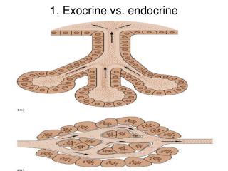
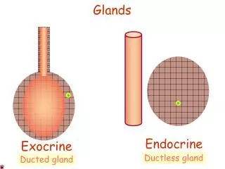
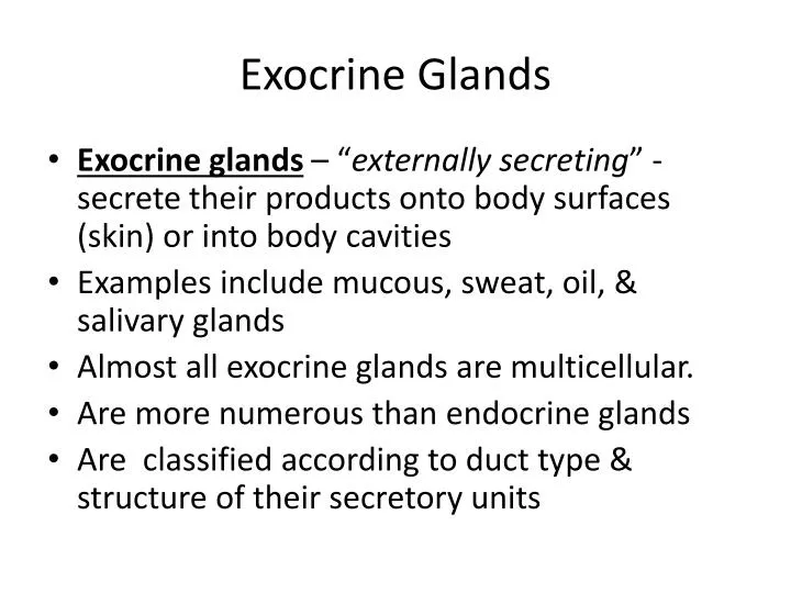
Exocrine Glands. Exocrine glands – “ externally secreting ” - s ecrete their products onto body surfaces (skin) or into body cavities Examples include mucous, sweat, oil, & salivary glands Almost all exocrine glands are multicellular. Are more numerous than endocrine glands

E N D
Exocrine Glands • Exocrine glands – “externally secreting” - secrete their products onto body surfaces (skin) or into body cavities • Examples include mucous, sweat, oil, & salivary glands • Almost all exocrine glands are multicellular. • Are more numerous than endocrine glands • Are classified according to duct type & structure of their secretory units
Functions of Connective Tissue • Functions of Connective tissue • 1- Binding & support • 2 - Protection • 3 - Insulation • 4 - Transportation
Characteristics of Connective Tissue • Characteristics of connective tissues • 1 – All connective tissues arise from mesenchyme –(an embryonic tissue) so they all have a common tissue of origin • 2 – Connective tissue has varying degrees of vascularity (amounts of blood vessels running through them) • ex: cartilage has few blood vessels, bone has more • 3 - Nonliving extracellular matrix, consisting of ground substance and fibers
Characteristics of Connective Tissue 3 – Connective tissue is composed mostly of nonliving extracellular matrix, consisting of ground substance & fibers which separates, often widely, the living cells of the tissue. (Other primary tissues are mostly composed of cells)
Types of Connective Tissue Proper • 1 – Loose Connective Tissue • 1) Areolar connective tissue - gel-like matrix with all three connective tissue fibers (collagen, elastic, & reticular) • serves to bind body parts together while allowing them to move freely over one another • wraps small blood vessels & nerves, surrounds glands, & cushions organs • is widely distributed throughout body
Connective Tissue Proper: Loose Figure 4.9a
Connective Tissue Proper: Loose • 2) Adipose connective tissue – (fat) richly vasculatized tissue that is similar to areolar connective tissue with closely packed adipocytes • Functions: • 1- reserves food stores • 2- insulates against heat loss • 3- supports • 4 - protects
Connective Tissue Proper: Loose • Found under skin, around kidneys, within abdomen, & in breasts • Local fat deposits serve nutrient needs of highly active organs
Connective Tissue Proper: Loose Figure 4.9b
Connective Tissue Proper: Loose • 3) Reticular connective tissue – loose ground substance with reticular fibers • Reticular cells lie in a fiber network • Forms a soft internal skeleton, or stroma, that supports other cell types • Found in lymph nodes, bone marrow, & spleen
Connective Tissue Proper: Loose Figure 4.9c
Types of Dense Connective Tissue • 2 - Dense Connective Tissue - all have fibers • Also called fibrous connective tissues • 1) Dense Regular connective tissue - contain closely packed bundles of parallel collagen fibers (running in same direction) with a few elastic fibers • Makes up tendons & ligaments
Connective Tissue Proper: Dense Regular Figure 4.9d
Types of Dense Connective Tissue • 2) Dense Irregular Connective Tissue – contains thick bundles of collagen fibers arranged in an irregular way with some elastic fibers • can withstand tension in many directions providing structural strength • is found in dermis & submucosa of digestive tract
Connective Tissue Proper: Dense Irregular Figure 4.9e
Types of Cartilage • Types of Cartilage • 1- Hyaline Cartilage – (gristle) most abundant cartilage in body • Matrix has network of collagen fibers • Functions: 1) supports 2) reinforces 3) cushions & 4) resists compression • **provides firm support with some pliability • Found in embryonic skeleton, end of long bones, nose, trachea, & larynx
Connective Tissue: Hyaline Cartilage Figure 4.9f
Types of Cartilage • 2- Elastic Cartilage – nearly identical to hyaline cartilage but with more elastic fibers • Maintains shape & structure while allowing flexibility • Supports external ear (pinna) & epiglottis
Connective Tissue: Elastic Cartilage Figure 4.9g
Types of Cartilage • 2- Fibrocartilage – structuaral intermediate between hyaline cartilage & regular connective tissues & has matrix similar to hyaline cartilage but less firm with thick collagen fibers • found where strong support & the ability to withstand heavy pressure are required such as the intervertebral discs & in discs of knee joint • provides tensile strength & absorbs compression shock
Connective Tissue: Fibrocartilage Cartilage Figure 4.9h
Connective Tissue: Bone (Osseous Tissue) • Bone - hard, calcified matrix with collagen fibers • also called osseous tissue • Osteocytes (mature bone cells)are found in small spaces called lacunae & are well vascularized with blood vessels • has exceptional ability to support & protect body structures due to its hardness, which is determined by collagen fibers & calcium salts found in extracellular matrix
Connective Tissue: Bone (Osseous Tissue) Figure 4.9i
Connective Tissue: Blood • Blood – consists of red & white blood cells & plasma proteins in a fluid matrix (plasma) • contained within blood vessels • f unctions in the transport of respiratory gases, nutrients, & wastes • is classified as a connective tissue because it developed from mesenchyme
Connective Tissue: Blood Figure 4.9j
Nervous Tissue • Nervous Tissue – main componenet of nervous system • Neurons – highly specialized branched nerve cells with long cellular processes that generate & conduct nerve impulses • dendrites – respond to stimuli • axons – carry impulses away from nerve cell body • support cells • transmits electrical signals from sensory receptors to effectors • Found in the brain, spinal cord, and peripheral nerves
Nervous Tissue Support cells – nonconducting cells that support, insulate, & protect delicate neurons • Neurons are found in brain, spinal cord, & peripheral nerves
Nervous Tissue Figure 4.10
Types of Muscle Tissue • Muscle Tissue – highly cellular, well-vascularized • responsible for most types of body movement • 1- Skeletal Muscle - attaches to bones of skeleton • Forms flesh of body & cause voluntary movement • Skeletal muscle cells (called muscle fibers) are long, cylindrical, multinucleate cells with striations
Muscle Tissue: Skeletal Figure 4.11a
Types of Muscle Tissue • 2- Cardiac Muscle - branching, striated, uninucleate cells interlocking at intercalated discs • responsible for involuntary movement of heart • 3- Smooth Muscle – has no striations • propels substances along internal passageways (i.e., peristalsis) • found in walls of hollow organs • is involuntary muscle
Muscle Tissue: Cardiac Figure 4.11b
Muscle Tissue: Smooth Figure 4.11c
Coverings & Lining Membranes • Some of the body’s membranes incorporate more than 1 type of tissue. • 1 – Cutaneous Membrane - cutis = skin • Consists of stratified squamous epithelium firmly attached to a thick layer of dense irregular connective tissue (dermis)
Coverings & Lining Membranes • 2- Mucous Membrane– also called mucosae • line body cavities such as those of hollow organs of digestive, respiratory, & urogenital tracts that open to exterior & contain either stratified squamous or simple columnar epithelia
Coverings & Lining Membranes • 3 – Serous Membrane – moist membranes found in closed ventral body cavities • consists of stratified squamous epithelium resting on a thin layer of loose connective (areolar) tissue • Are named by where they are located • Pleura = lungs • Pericardium = covering of heart • Peritoneums = coverings in abdominopelvic cavity
Tissue Repair • Steps of Tissue Repair– • 1- Inflammation – caused by tissue trauma & is characterized by dilation of blood vessels, increase in vessel permeability, redness, heat, swelling, & pain • Blood clot forms
Tissue Repair • 2 – Organization restored the blood supply. • Blood clot is replaced with granulation tissue • 3 – Regeneration & Fibrosis • If wound is small & damaged tissue can divide, the tissue will regenerate & cover the fibrous tissue • When wound is extensive or damaged tissue cannot divide, it is repaired only by fibrous connective (scar) tissue.
Developmental Aspects • 3 primary germ layers for early during embryonic development & becomes specialized into the 4 types of tissues. • Ectoderm – top layer - nervous tissue comes from ectoderm • Mesoderm – middle layer – muscle & connective tissue come from mesoderm • Endoderm – inside layer – organs • Epithelium arises from all 3 primary germ layers