Download

1 / 23
250 likes | 667 Vues
Lecture 4 Beam restricting Devices, their Use and Maintenance. Topic 1 Importance of beam restricting devices Topic 2 Types of beam restricting devices Topic 3 Quality assurance tests of beam restricting devices Topic 4 Compression of area of interest.

E N D
Lecture 4 Beam restricting Devices, their Use and Maintenance • Topic 1 Importance of beam restricting devices • Topic 2 Types of beam restricting devices • Topic 3 Quality assurance tests of beam restricting devices • Topic 4 Compression of area of interest
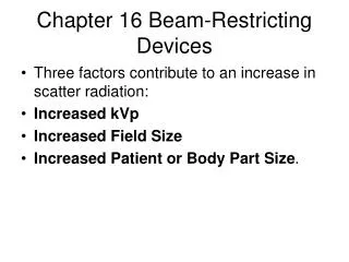
Topic 1 Importance of beam restricting devices • These devices restrict the primary x-ray beam to the area of interest. • The larger the area covered by primary x-ray beam, the greater the scattered radiation produced. • Scattered radiation must be minimized. They increase radiation dose and add to film darkening thus reducing visualization of details.
Topic 1 Importance of beam- restricting devices Beam was not restricted to the chest – the area of interest. Entire abdomen and part of face of baby was exposed unnecessarily.
Topic 1 Importance of beam- restricting devices Unrestricted beam-considerable scatter Restricted beam – less scattered radiation
Topic 2 Types of beam-restrictingdevices a. Attached to x-ray tube housing • Aperture diaphragms - lead sheets with circular, square, or rectangular openings • Cones – detachable metal tubes of different lengths, shapes, and sizes of opening • Variable aperture collimators – adjustable lead plates or shutters , contain "cross hairs", a light source, and a mirror to project the light
Beam- restricting devices Removable metal cone Variable aperture collimator
Variable aperture, multiple shutter collimator Diaphragm closer to window reduces off-focus x rays. Multiple shutters Multiple shutters Smaller sized field reduces size of primary beam.
Topic 2 Types of beam - restricting devices b. Not attached to x ray tube housing Lead blockers – used to divide a cassette when more than one exposure is made on a single film; reduces unnecessary exposure of film thus improving image quality
Use of lead blockers • Place blocker on couch for lateral projections of thoracic, lumbar, sacrum or coccyx spine. • Position blocker next to patient's back to absorb radiation that will not pass through spine. • Defective lead rubber aprons could be cut into a range of sizes to serve as blockers.
Topic 3 Quality assurance tests of beam limiting devices • Collimation or limiting the x-ray beam to the area of interest requires use of beam restricting devices. It is good practice to collimate such that the resulting image has collimated edges on all four sides of film
Topic 3 Quality assurance tests of beam- restricting devices • Bring tube to rest on table top to check • that all four edges of square light beam • diaphragm touch the table top when • using vertical beam. Do several times • during the day. • Do the same test with cone inserted in • front of the x ray tube window. • Caution: Cones sometimes become bent when dropped or bumped.
Topic 3 Quality Assurance tests of beam restricting devices • Do collimator-beam alignment check at least every six months. • Do check of alignment of centre of x ray beam at least every six months.
Collimator-beam alignment test • Using 100 cm focus-film distance (FFD), set collimator at 20 cm x 20 cm field size on surface of loaded cassette. • Place metal coins or paper clips on top of cassette as shown in next slide. • Expose the film at 60 kVp and 4 – 8 mAs. • Process the film. • Check whether the distance between outer edges of image and of paper clips/coins is within +/- 2% of FFD.
Line diagram showing metal coins and field size Coin to identify right upper corner. Edge of light. Cassette.
Check of alignment of centre of x-ray beam • Place unexposed loaded cassette in centre of bucky tray and centre tube to cassette. • Move tube to 100 cm FFD to bucky tray. Reduce longitudinal collimators to a thin slit (e.g. 0.5 cm). Close lateral collimators. Expose using 60 kVp and 4 – 8 mAs. • Do not remove cassette.
Check of alignment of centre of x ray beam (cont) • Close longitudinal collimators and open lateral collimators to a thin slit. Expose film again. Process film. • Bend film in half and check that exposed "cross" is in film centre. • Acceptable deviation: 1 cm on either side of centre. • Can also be done for non-bucky radiography. Just move tube so 100 cm FFD is to cassette top.
Topic 3 Summary Beam restricting devices • may be attached or not attached to tube housing • require periodic checks • are necessary for radiation protection and contribute to good image quality • minimize primary radiation • minimize scatter radiation
Topic 4 Compression of area of interest • Reducing patient thickness by compression minimizes production of scattered radiation, thus reducing radiation dose. • Device used : compression band.
Topic 4 Compression of area of interest Compression band over abdomen reduces its volume. This reduces scatter radiation.
Lecture 4 Summary • Beam-restricting devices are important in radiation protection. • These devices should be checked regularly. • Regular performance of quality assurance tests is in accordance with ALARA.