Download
1 / 34
360 likes | 590 Vues
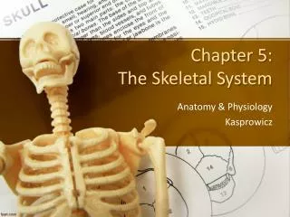
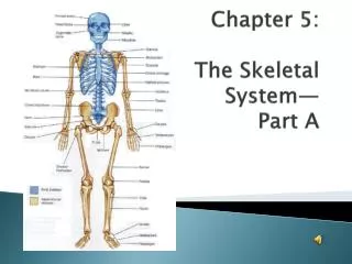
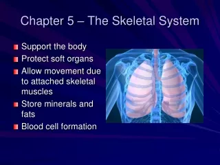
Chapter 5 Skeletal System. Skeleton. Greek meaning “dried up body” Axial & Appendicular Functions: Support Protection Movement Storage Blood cell formation. Bones. 206 bones Compact and spongy bone Long bones Short bones Flat bones Irregular bones. Long Bones Gross Anatomy.

E N D
Skeleton • Greek meaning “dried up body” • Axial & Appendicular • Functions: • Support • Protection • Movement • Storage • Blood cell formation
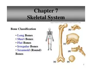
Bones • 206 bones • Compact and spongy bone • Long bones • Short bones • Flat bones • Irregular bones
Long BonesGross Anatomy • Diaphysis – compact bone • Periosteum – covering diaphysis • Epiphyses – ends • Articular cartilage – covers epiphyses • Epiphyseal line – remnant of plate • Yellow marrow – fat storage • Red marrow – RBCs • Bone markings (table 5.1)
Long BonesMicroscopic Anatomy • Osteocytes – mature bone cells • Lacunae – cavities • Lamellae – circles of lacunae • Haversiancanals – cavity with blood supply • Osteon – canal and matrix rings • Canaliculi – connect cells to haversian canals • Perforatingcanals – canals at right angles
Bone Formation, Growth and Remodeling • Ossification has two major phases • Hyaline model is covered with bone cells (osteoblasts) • Hyaline model digested away, making medullary cavity. Hyaline stays in the articular cartilages and epiphyseal plates Bone remodeling: Osteoclasts – activated by PTH to destroy bone and release calcium into the blood stream
Homeostatic Imbalance • Rickets: • Childhood disease in which bones fail to calcify. Lack of vitamin D or of calcium. Milk, bread and others are fortified with Vit D. Sun.
Bone Fractures • Simple fracture – bone breaks cleanly and does not penetrate skin • Compound fracture – broken bone ends penetrate skin • Fracture treated by reduction • Closed reduction: bone ends are put back into normal position by doctor • Open reduction: surgery is performed to secure them with pins/plates/wires
Compound Fracture Open Reduction
Fracture Healing • Four events: • 1. Hematoma formed by ruptured blood vessels • 2. Break splinted by fibrocartilage callus (cartilage matrix, bony matrix and collagen fibers). “Splints” the bone, closing the gap • 3. Bony callus formed (spongy bone) • 4. Bony callus remodeled over months time
Axial Skeleton • Skull: • Cranium and Facial Bones • Cranium bones: • Frontal (forehead) • Parietal (superior & lateral. Paired… form sagittal suture and coronal suture) • Temporal (inferior to parietal… form squamous sutures. External auditory meatus, styloid process, zygomatic process, mastoid process, jugular foramen) • Occipital (most posterior. Forms lambdoid suture. Opening called foramen magnum. Occipital condyles) • Sphenoid (floor of cranial cavity. Sella turcica, foramen ovale, sphenoid sinuses) • Ethmoid (roof of nasal cavity. Crista galli, cribiform plates) • Facial Bones: • Maxillae (paired and fuse to form upper jaw. Palantine process, paranasal sinuses) • Palantine (paired. Posterior part of hard palate. Cleft palate) • Zygomatic (paired. Cheekbones) • Lacrimal (paired. Medial walls of orbit) • Nasal (paired. Bridge of nose) • Vomer (single bone. Nasal septum) • Inferior Conchae (paired. Inside nasal cavity) • Mandible (single bone. Largest, strongest bone in face) • Hyoid Bone • Closely related to mandible and temporal bones. Suspended midneck above larynx. Movable base for tongue and attaches neck muscles)
Axial Skeleton • Vertebral Column (spine) • Intervertebral discs • 24 bones • 7 cervical (neck) • Atlas and axis • 12 thoracic • 5 lumbar • Sacrum • 5 Fused vertebrae • Coccyx • 3-5 fused tiny vertebrae. “tailbone”
Vertebrae • Body • Vertebral arch • Vertebral foramen • Transverse processes • Spinous process • Superior and inferior articular processes
Axial Skeleton • Bony Thorax • Sternum • Jugular notch, sternal angle, xiphisternal joint • Ribs • 12 pairs. True ribs (1st 7), False ribs (last 5 NOT attached to sternum)
Appendicular Skeleton • Shoulder Girdle • Upper Limbs • Pelvic Girdle • Lower Limbs
Shoulder Girdle • Clavicle • Scapulae • Acromion • Coracoid process • Glenoid cavity
Bones of the Arm • Humerus • Greater, lesser tubercles • Deltoid tuberosity • Radial groove • Trochlea • Capitulum • Radius – lateral • Radioulnar joints • Interosseous membrane • Radial tuberosity • Ulna – medial • Coronoid process • Olecranon process • Trochlear notch • Carpal bones • Carpals • Metacarpals • Phalanges
Greater tubercle Trochlear notch Lesser tubercle Olecranon process Coronoid process Radial tuberosity Proximal radioulnar joint Radial groove Deltoid tuberosity Interosseous membrane Ulna Radius Distal radioulnar joint Capitulum Trochlea Posterior Humerus Anterior Humerus
Pelvic Girdle • Ilium • Sacroiliac joint • Iliac crest • Ischium • Pubis • Pubic symphysis • Acetabulum sacroiliac joint iliac crest ilium sacrum pubic bone acetabulum ischium Pubic symphysis
Lower Limbs • Thigh • Femur • Greater, lesser trochanters • Intertrochanteric line/crest • Gluteal tuberosity • intercondylar notch • Leg • Tibia (larger, more medial) • Tibial tuberosity • Medial malleolus • Anterior crest • Fibula • Lateral malleolus • Foot • Tarsus (7 tarsal bones) • Calcaneus • Talus • Metatarsals • 14 phalanges
Joints • Functionally • Synarthroses (immovable) • Amphiarthroses (slightly movable) • Diarthroses (freely movable) • Structurally • Fibrous • Cartilaginous • Synovial
Fibrous Joints • Sutures of skull • Syndesmoses (tibia and fibula)
Cartilaginous Joints • Pubic symphysis • Intervertebral joints • Hyaline-cartilage epiphyseal pates of growing long bones • Cartilaginous joints between first ribs/sternum
Synovial Joints • Four characteristics • Articular cartilage • Fibrous articular capsule • Joint cavity • Reinforcing ligaments • ASSOCIATED… • Bursae • Tendon sheath
Synovial Joints Based on Shape • Plane Joint • Nonaxial • Intercarpal joints of wrist • Hinge Joint • Uniaxial • Elbow, ankle, joints between phalanges • Pivot Joint • Uniaxial • Proximal radioulnar joint • Condyloid joint • “knuckle-like”, biaxial • Metacarpophalangeal joints • Saddle joint • Biaxial • Carpometacarpal joints in thumb • Ball-and-socket joint • Multiaxial • Shoulder, hip
Inflammatory Disease • Bursitis • Sprain • Arthritis • Osteoarthritis • Rheumatoid arthritis • Gouty arthritis
Developmental Aspects • Birth: UL ratio 1.7:1 • 10 yo: UL ratio 1:1 • Osteoporosis