Download

1 / 44
680 likes | 1.83k Vues
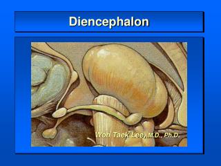
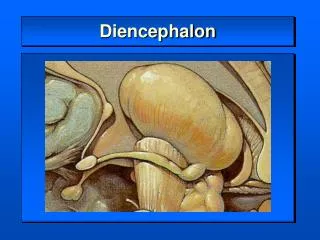
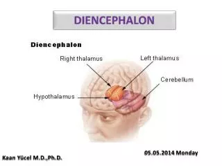
DIENCEPHALON. Dr. Zeenat Zaidi. Diencephalon. Paired structure Located between the brain stem and the cerebral hemisphere Continuous with the rostral part of the midbrain Forms the lateral wall of the 3 rd ventricle. mb. p. C. mo.

E N D
DIENCEPHALON Dr. Zeenat Zaidi
Diencephalon • Paired structure • Located between the brain stem and the cerebral hemisphere • Continuous with the rostral part of the midbrain • Forms the lateral wall of the 3rd ventricle mb p C mo
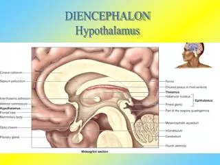
Almost entirely surrounded by the cerebral hemispheres A little part seen externally on the base of the brain caudal to optic chiasma, includes: Infundibulum Tuber cinerium Mamillary bodies Other parts seen on sagittal & coronal sections tc I mb
Fornix CC Dorsal Ventral Midbrain Optic chiasma Cerebral aqueduct • On the medial surface, the diencephalon is subdivided, by hypothalamic sulcus (indicated by black line) into: • Dorsal part • Ventral part
Dorsal part Thalamus &Epithalamus Subthalamus & Hypothalamus Ventral part H
Relations Lateral: Internal capsule Dorsal: Lateral ventricle Medial: 3rd ventricle Ventral: Exposed on the base of the brain
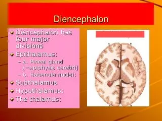
Epithalamus • Relatively small part, located in most caudal and dorsal region • Lies immediately rostral to superior colliculus • Consists of: • Pineal gland & • Habenular nuclei
Pineal Gland • An endocrine organ • Synthesizes melatonin • Controls: • Sleep/awake cycle • Regulation of onset of puberty
Habenular nuclei • Located in habenular triangle (area in the posterior part of the diencephalon, just anterior to pineal gland) • Have connections with limbic system • Serves autonomic function and emotional drives
Thalamus • Large mass of grey matter, in shape and size, resembles small hen’s egg • Forms the lateral wall of the 3rd ventricle • Separated from hypothalamus by hypothalamic sulcus • May be connected to opposite thalamus by interthalamic adhesion (massa intermedia) thalamus Interthalamic adhesion Hypothalamic sulcus
Thalamus: In horizontal sections of brain Higher level Lower level
Thalamus has a narrow anterior end called tubercle of thalamus, that lies in the posterior boundary of the interventricular foramen The expanded posterior end, called pulvinar, lies above the superior colliculi
Relations Dorsal: lateral ventricle Anterior: interventricular foramen Medial: 3rd ventricle Lateral: Internal capsule Ventral: Subthalamus & Hypothalamus Caudal: midbrain
Surfaces • 4 Surfaces: • Superior • Inferior • Medial • Lateral S L M l
Superior Surface caudate nucleus stria terminalis • Bounded laterally by caudate nucleus, thalamostriate vein and a nerve fiber bundle called striaterminalis • Lateral part lies in the floor of the lateral ventricle & is covered by ependyma • Medial part is related to the choroid plexus of the 3rd ventricle LV thalamo-striate vein ependyma choroid plexus
Lateral Surface Related to the internal capsule Inferior Surface Rests on the subthalamus & hypothalamus
Medial Surface Stria medullaris thalami • Striamedullaris thalami (a fascicle of nerve fibers) courses along its dorsomedial margin • Below is limited by hypothalamic sulcus • Forms the upper part of the lateral wall of the 3rd ventricle • Covered by ependyma Hypothalamic sulcus
Internal Organization • Thalamus is composed of grey matter, interrupted by two vertical sheaths of white matter called medullarylaminae. • External medullary lamina: • Located laterally, separates reticular nucleus from the rest of the thalamic mass • Contains thalamocortical & corticothalamic fibers
Internal medullary lamina Y- shaped band, divides thalamusinto Anterior,Medial&Lateral nuclear groups Contains: Fibers connecting thalamic nuclei with one another Neuronal collections called intralaminar nuclei
Nuclear Groups • Anterior nuclear group • Lateral nuclear group: Divided into: • dorsal & • ventral tiers • Medial nuclear group • Intralaminar nuclei • Reticular nucleus • Midlinenuclei
Functional Organization • All the nuclei of the thalamus except reticular nucleus, project to ipsilateral cerebral cortex • The whole of the cerebral cortex receives input from the thalamus • All thalamic nuclei receive corticofugalfibersin a basically reciprocal fashion
Based on their connection with the cerebral cortex, the thalamic nuclei are divided into: • Specific nuclei • Nonspecific nuclei
Specific nuclei: • Have well-defined sensory and motor functions • Have highly organized point-to-point connection with sensory & motor regions of cerebral cortex • Lie within the ventral group of the lateral nuclear group • Non-specific Nuclei: • Receive less functionally distinct afferent input • Connect with wider area of cortex, including associative and limbic regions • Include nuclei of the dorsal tier of lateral group, and whole of the anterior and medial group
Lateral Nuclear GroupVentral Tier • Ventral anterior • Ventral lateral • Ventral posterior: • (VPL) • (VPM) • Lateral geniculate • Medial geniculate
Ventral Anterior Nucleus Influences motor activity Ipsilateralglobuspallidus& substantianigra Motor cortex Premotor & supplementary motor cortex
Ventral Lateral Nucleus Influences motor activity Ipsilateralglobuspallidus & substantianigra Contralateraldentate nucleus Primary motor cortex Primary motor cortex
Ventral Posterior Nucleus Chief sensory relay station General sensory afferents from the contralateral half of the Head& neck(VPM) and Body(VPL) Primary somatosensory cortex
Lateral Geniculate Body Part of the Visual Pathway Ipsilateraltemporal hemiretina Contralateralnasal hemiretina Optic radiation to the primary visual cortex
Medial Geniculate Body Part of the Auditory Pathway Inferior colliculus Auditory radiation to the primary auditory cortex
Lateral Nuclear GroupDorsal Tier • Lateral Dorsal • Lateral Posterior • Pulvinar
Lateral dorsal nucleus • Part of Limbic System • Hippocamus • Cingulategyrus Lateral posterior nucleus sensory association cortex of parietal lobe Pulvinar: Sensory association cortices of parietal, temporal & occipital lobes
Medial Nuclear Group • Integrates emotion, thought, and judgment Mediodorsal nucleus & Nucleus reuniens Hypothalamus, amygdala, other thalamic nuclei, prefrontal cortex Prefrontal cortex & limbic structures
Anterior Nuclear Groups Functionally part of the limbic system. Involved in control of instinctive drives, emotional aspect of behaviour and in memory • 3 parts: • Anteroventral • Anteromedial • Anterodorsal
Mammillary body of hypothalamus via mammillothalamic tract cingulate gyrus
Midline Nuclei Brainstem reticular formation Cingulate gyrus and hypothalamus • Located between medial nuclear group and the ependyma of 3rd ventricle • Important in visceral functions
Intralaminar Nuclei • Located within the internal medullary lamina • Main nuclei: Centromedian & Parafascicular • Function as activator of the cerebral cortical mantle • Lesions reduce the perception of pain and level of conciousness
Reticular formation, spinothalamic & trigeminothalamic systems Widespread regions of cerebral cortex, caudate & putamen of the basal ganglia
Reticular Nucleus Regulates the activity of thalamus • Located between the external medullary lamina & the internal capsule Collaterals of both Thalamocortical & Corticothalamic fibers Other thalamic nuclei
Functions of the thalamus • Receives and analyses all the sensory information (except olfactory) from the body • Having extensive connections with the basal ganglia and the motor cortices, it plays a pivot role in voluntary motor activity. Thalamotomy (VA, VL) was once used to treat basal ganglia disorders • Connections with the limbic system makes it important in the control of mood, emotional and sexual behavior, and memory
Thalamic Lesions • Cerebrovascular lesions or tumors of thalamus lead to: • Loss of sensation in the contralateral side of face and body followed by distressing discomfort, & burning and diffuse pain in the anaesthetic areas (thalamic pain) • Thalamic syndrome: Abnormal voluntary movements (chorea or hemiballismus) with hemisensory disturbance