Download
1 / 77
800 likes | 1.73k Vues
Diencephalon. By Rashid Alshahoumi. Overview Development of Diencephalon Basic Organization Dorsal Thalamus (Thalamus). Hypothalamus Ventral Thalamus ( Subthalamus) Epithalamus Vasculature of the Diencephalon. Outline:. Diencephalon. The diencephalon includes - Dorsal thalamus

E N D
Diencephalon By Rashid Alshahoumi
Overview Development of Diencephalon Basic Organization Dorsal Thalamus (Thalamus) Hypothalamus Ventral Thalamus ( Subthalamus) Epithalamus Vasculature of the Diencephalon Outline:
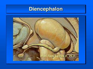
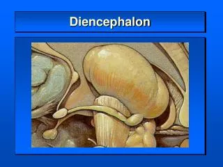
Diencephalon • The diencephalon includes - Dorsal thalamus - Hypothalamus - Ventral thalamus - Epithalamus • Situated between telencephalon & brainstem. • Main processing center for information
Rt & Lt halves of the diencephalon,contain symmetrically distributed cell groups separated by the space of the 3rd ventricle
Development of the Diencephalon • The cell groups that give rise to the diencephalon form in the caudomedial portion of the prosencephalon, bordering on the space that will become the 3rd ventricle. • The developing brain at this level consists initially of a roof plate and the two alar plates; it lacks a well-defined floor plate and basal plates.
The hypothalamic sulcus • A shallow groove appears in the wall of the 3rd ventricle & extends rostrally from the developing cerebral aqueduct to the ventral edge of the interventricular foramen • Divides the alar plate into : • Superior (dorsal) area : future dorsal thalamus • Inferior (ventral) portion : future hypothalamus • The dorsal thalamus - On each side of the 3rd ventricle increases rapidly in size & will partially fuse across the space of the 3rd ventricle to form : - massaintermedia, or interthalamic adhesion . (present in about 80% of the general population)
The epithalamus - Develops from the caudal portion of the roof plate. - By 7th week, a small thickening of the roof plate forms. It gradually increases in size & evaginates to form the epiphysis, which develops into the pineal gland of the adult . - The portion of the roof plate immediately rostral to the epiphysis gives rise to the habenula, a small thickening in which the habenular nuclei will develop • Just anterior to the habenular region, the roof plate epithelium & adjacent pia mater give rise to the choroid plexus of the third ventricle, This choroid plexus is continuous through the interventricular foramina with that of the lateral ventricles.
In locations around the perimeter of the 3rd ventricle, specialized patches of ependyma lie on the midline & form unpaired structures called the circumventricular organs • These structures include : • Subfornical organ • Organum vasculosum of the lamina terminalis • Subcommissural organ, • Pineal gland.
The development of the pituitary gland during the 3rd week is linked to that of the diencephalon . • A downward extension of the floor of the 3rd ventricle, the infundibulum, meets the Rathke pouch, an upward outpocketing of the stomodeum, the primitive oral cavity.
By the end of the 2nd month, the Rathke pouch loses its connection with the developing oral cavity but maintains its attachment to the infundibulum. • As development continues, the Rathke pouch gives rise to the anterior lobe (adenohypophysis) and pars intermedia of the pituitary gland • Infundibulum differentiates into the posterior lobe of the pituitary gland, or neurohypophysis
A craniopharyngioma (Rathke pouch tumor) can arise from a portion of the Rathke pouch that fails to undergo proper migration & apposition to the infundibulum. • These tumors mimic lesions of the pituitary & may cause visual problems, diabetes insipidus, & ↑ ICP
Basic Organization • The junction between the diencephalon & midbrain lies along a line extending from the posterior commissure to the caudal edge of the mammillary body on the medial aspect of the hemisphere . • On the surface of the hemisphere, this interface is represented by a line starting at the caudal aspect of the mammillary body, extending anterolaterally over the edge of the crus cerebri & following the caudal edge of the optic tract . • The boundary between the diencephalon & surrounding telencephalon is less distinct & represented : - laterally by the internal capsule - rostrally by the interventricular foramen, lamina terminalis & optic chiasm .
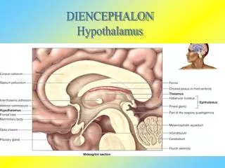
The 3rd third ventricle has small evaginations or recesses associated with • Optic chiasm (supraoptic recess) • Infundibulum (infundibular recess) • Pineal gland (pineal& suprapineal recesses)
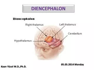
The dorsal thalamus • Located superior to the hypothalamic sulcus • Extends from the interventricular foramen caudally to the level of the splenium of the corpus callosum. • The hypothalamus • Located inferior to the hypothalamic sulcus • Bordered : - Rostrally by the lamina terminalis - Caudally by a line that extends from the posterior aspect of the mammillary body - Superiorly to intersect with the hypothalamic sulcus.
The ventral thalamus (subthalamus) • Located : • Caudal to the hypothalamus • Rostral to the diencephalon-midbrain junction • Lateral to the midline • Epithalamic structures • Located posteriorly & caudally in close apposition to the posterior commissure • Include : - pineal gland - habenular nuclei - main afferent bundle of these nuclei - stria medullaris thalami .
Dorsal Thalamus (Thalamus) • The dorsal thalamus (or thalamus) is a massive collection of neuronal cell groups that participate in a widely diverse array of functions involving motor, sensory & limbic systems. • Typically, thalamic output neurons project to the cerebral cortex → !! very little information reaches the cerebral cortex without first being processed by thalamic neurons→ the thalamus is functional "gateway" to the cerebral cortex
In turn, nearly all regions of the cerebral cortex give rise to reciprocal projections that return to the thalamic region from which they originally received input.
The thalamus is covered on its lateral aspect by a layer of myelinated axons, the external medullary lamina ( includes fibers that enter or leave the subcortical white matter) • Within the external medullary lamina are clusters of neurons that form the thalamic reticular nucleus.
An internal medullary lamina: - Consisting of myelinated fibers - Extends into the substance of the thalamus, where it forms partitions or boundaries that divide the thalamus into its principal cell groups : - anterior, medial, lateral &intraluminar nuclear groups. • There are midline thalamic nuclei located just superior to the hypothalamic sulcus. • Finally, attached to the caudolateral portion of the thalamus are the medial and lateral geniculate bodies (and their nuclei) .
Anterior Thalamic Nuclei • This group of cells consists of a large principal nucleus & two smaller nuclei → form the anterior nucleus of the thalamus • The anterior nucleus forms a prominent wedge on the rostral aspect of the dorsal thalamus just caudolateral to the interventricular foramen → this wedge is the anterior thalamic tubercle. • Rostrally, the internal medullary lamina divides to partially encapsulate the anterior nucleus.
Anterior Thalamic Nuclei • The cells of this nucleus receive dense limbic-related projections from (1) the mammillary nuclei via the mammillothalamic tract and (2) the medial temporal lobe (hippocampus) via the fornix. • The output of this nucleus is primarily directed to the cingulate gyrus through the anterior limb of the internal capsule.
Medial Thalamic Nuclei • Comprises the dorsomedial nucleus • Composed of : -Large parvicellular (located caudally) -Magnocellular (located rostrally) -Small paralaminar adjacent to the internal medullary lamina • 2 larger portions are linked to parts of the frontal & temporal lobes & to the amygdaloid complex . • Cells of the paralaminar subdivision receive input from the frontal lobe & substantia nigra (may play a role in the control of eye movement)
Lateral Thalamic Nuclei 2 subdivisions • Dorsal subdivision • Lateral dorsal - Functionally part of anterior group (limbic system) • Lateral posterior
Inputs Pretectal area Superior colliculus Roles: Visual relay center Selective attention Speech Pulvinar Border with lateral posterior is vague Reciprocal connections: Lateral geniculate nucleus Parietal lobe Temporal lobe Occipital lobe
Ventral subdivision - Ventral anterior - Ventral lateral - Ventral posterior • Receive direct input from long ascending tracts • Reprocal connections with cortex • Retrograde degeneration on cortical lesions
Input Globus pallidus Substantia nigra Intralaminar nucleus (thalamus) Premotor/prefrontal cortex Output (reciprocal connections) : Premotor cortex Prefrontal cortex Intralaminar nucleus Roles: Motor relay station - Regulate movement (Control of voluntary movement) Medial part - Eye, head, neck Lateral part - Body, limb Ventral Anterior
Input Deep cerebellar nuclei Globus pallidus Primary motor cortex Output Primary motor cortex (reciprocal) Parietal lobe -Somatosensory areas Premotor/Supplementary motor areas Role: Motor relay station - Cerebellum/basal ganglia/cortex Ventral Lateral Nucleus
2 Divisions Ventral posterior medial (VPM) Ventral posterior lateral (VPL) Inputs Medial lemniscus - VPL Spinothalamic -VPL Trigeminal lemniscus (taste) - VPM Primary somatosensory cortex - VPM & VPL Output Primary somatosensory cortex (reciprocal) Parietal operculum (taste) Ventral Posterior Nucleus
The lateral (LGB) and medial (MGB) geniculate nuclei are considered parts of the lateral thalamic nuclear group . • MGB receives ascending auditory input via the brachium of the inferior colliculus → projects to the primary auditory cortex in the temporal lobe. • LGB receivesvisual input from the retina via the optic tract → projects to the primary visual cortex on the medial surface of the occipital lobe .
Located in the posterior thalamus at about the level of the pulvinar and geniculate nuclei is a cluster of cell groups collectively called the posterior nuclear complex. • This complex consists of : - Suprageniculate nucleus • Nucleus limitans • Posterior nucleus • These nuclei are positioned superior to the medial geniculate and medial to the rostral pulvinar. • The posterior nuclear complex receives& sends to the cortex nociceptive cutaneous input that is transmitted over somatosensory pathways
Intralaminar Nuclei • Embedded within the internal medullary lamina are the discontinuous groups of neurons that form the intralaminar nuclei. • Projections to the neostriatum & to other thalamic nuclei, along with diffuse projections to the cerebral cortex. • 2 of the most prominent cell groups are : • Centromedian : projects to the neostriatum & to motor areas of the cerebral cortex • Parafascicular nuclei : projects to rostral & lateral areas of the frontal lobe. • Other intralaminar nuclei receive input from ascending pain pathways and project to somatosensory and parietal cortex.
Midline Nuclei • The midline nuclei are the least understood components of the thalamus?? • The largest is the paratenial nucleus, which is located just ventral to the rostral portion of the stria medullaris thalami; other cells are associated with the interthalamic adhesion (massa intermedia). • Inputs are poorly defined • Efferent fibers reach the amygdaloid complex &the anterior cingulate cortex, suggesting a role in the limbic system.
Thalamic Reticular Nucleus • The cells are situated within the external medullary lamina & between this lamina and the internal capsule . • Axons of these cells project medially into the nuclei of the dorsal thalamus or to other parts of the reticular nucleus, but not into the cerebral cortex. • Afferents are received from the cortex and from nuclei of the dorsal thalamus via collaterals of thalamocortical & corticothalamic axons. • Thalamic reticular neurons modulate, or gate, the responses of thalamic neurons to incoming cerebral cortical input .
Thalamic nucleus : efferent projections (thalamocortical axons) → corterx • Cortex → reciprocal projection (corticothalamic axons) → thalamic nucleus • VL/motor/precentral gyrus and anterior paracentral gyrus • VPL/sensory for the body/postcentral gyrus and posterior paracentral gyrus • VPM/sensory for the face/postcentral gyrus • MGB/auditory/transverse temporal gyrus • LGB/vision/cortex on the calcarine sulcus • The anterior nucleus projects primarily to the cingulate gyrus and functions in the broad area of behavior
Thalamic nuclei : relay nuclei or association nuclei • Thalamic nuclei : specific or nonspecific
Hypothalamus • The hypothalamus is mainly involved in visceromotor, viscerosensory &endocrine activities. • The hypothalamus & related limbic structures receive sensory input regarding the internal environment & in turn, regulate through four mechanisms the motor systems that modify the internal environment.
Is a principal modulator of autonomic nervous system function. Is a viscerosensory transducer, containing neurons with specialized receptors capable of responding to changes in the temperature or osmolality of blood, as well as to specific hormonal levels in the general circulation. It regulates the activity of the anterior pituitary through the production of releasing factors (hormone-releasing hormones) It performs an endocrine function by producing & releasing oxytocin &vasopressin into the general circulation within the posterior pituitary. Hypothalamus
The hypothalamus can be divided into lateral, medial & periventricular zones :
Lateral Hypothalamic Zone • Composed of diffuse clusters of neurons intermingled with longitudinally oriented axon bundles • Cells are involved in cardiovascular function & in the regulation of food & water intake.