Download
1 / 31
320 likes | 691 Vues
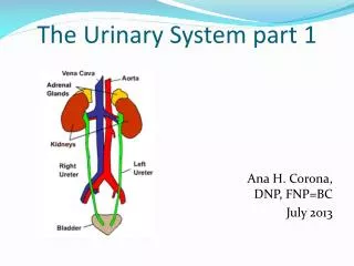
The Urinary System Part A. 25. Urinary System Organs. Kidneys – Urinary bladder – provides a temporary storage reservoir for urine Paired ureters – transport urine from the kidneys to the bladder Urethra – transports urine from the bladder out of the body. Urinary System Organs.

E N D
The Urinary System Part A 25
Urinary System Organs • Kidneys – • Urinary bladder – provides a temporary storage reservoir for urine • Paired ureters – transport urine from the kidneys to the bladder • Urethra – transports urine from the bladder out of the body
Urinary System Organs Figure 25.1a
Kidney Location and External Anatomy • The bean-shaped kidneys lie in a retroperitoneal position in the superior lumbar region and extend from the twelfth thoracic to the third lumbar vertebrae • The right kidney is lower than the left because it is crowded by the liver • The lateral surface is convex and the medial surface is concave, with a vertical cleft called the renal hilus leading to the renal sinus • Ureters, renal blood vessels, lymphatics, and nerves enter and exit at the hilus
Layers of Tissue Supporting the Kidney • Renal capsule – fibrous capsule that prevents kidney infection • Adipose capsule – fatty mass that cushions the kidney and helps attach it to the body wall • Renal fascia – outer layer of dense fibrous connective tissue that anchors the kidney
Kidney Location and External Anatomy Figure 25.2a
Internal Anatomy • A frontal section shows three distinct regions • Cortex – the light colored, granular superficial region • Medulla – exhibits cone-shaped medullary (renal) pyramids • Pyramids are made up of parallel bundles of urine-collecting tubules • Renal columns are inward extensions of cortical tissue that separate the pyramids • The medullary pyramid and its surrounding capsule constitute a lobe
Internal Anatomy • Renal pelvis – flat, funnel-shaped tube lateral to the hilus within the renal sinus
Internal Anatomy • Major calyces – large branches of the renal pelvis • Collect urine draining from papillae • Empty urine into the pelvis • Urine flows through the pelvis and ureters to the bladder
Internal Anatomy Figure 25.3b
Blood and Nerve Supply • Approximately one-fourth (1200 ml) of systemic cardiac output flows through the kidneys each minute • Arterial flow into and venous flow out of the kidneys follow similar paths • The nerve supply is via the renal plexus Figure 25.3c
The Nephron • Nephrons are the structural and functional units that form urine, consisting of: • Glomerulus – a tuft of capillaries associated with a renal tubule • Glomerular (Bowman’s) capsule – blind, cup-shaped end of a renal tubule that completely surrounds the glomerulus
The Nephron • Renal corpuscle – the glomerulus and its Bowman’s capsule • Glomerular endothelium – fenestrated epithelium that allows solute-rich, virtually protein-free filtrate to pass from the blood into the glomerular capsule
The Nephron Figure 25.4b
Anatomy of the Glomerular Capsule • The external parietal layer is a structural layer • The visceral layer consists of modified, branching epithelial podocytes • Extensions of the octopus-like podocytes terminate in foot processes • Filtration slits – openings between the foot processes that allow filtrate to pass into the capsular space
Renal Tubule • Proximal convoluted tubule (PCT) – composed of cuboidal cells with numerous microvilli and mitochondria • Reabsorbs water and solutes from filtrate and secretes substances into it
Renal Tubule • Loop of Henle – a hairpin-shaped loop of the renal tubule • Proximal part is similar to the proximal convoluted tubule • Proximal part is followed by the thin segment (simple squamous cells) and the thick segment (cuboidal to columnar cells) • Distal convoluted tubule (DCT) – cuboidal cells without microvilli that function more in secretion than reabsorption
Renal Tubule Figure 25.4b
Connecting Tubules • The distal portion of the distal convoluted tubule nearer to the collecting ducts
Connecting Tubules • Two important cell types are found here • Intercalated cells • Cuboidal cells with microvilli • Function in maintaining the acid-base balance of the body • Principal cells • Cuboidal cells without microvilli • Help maintain the body’s water and salt balance
Nephron type • Cortical nephrons – 85% of nephrons; located in the cortex • Juxtamedullary nephrons: • Are located at the cortex-medulla junction • Have loops of Henle that deeply invade the medulla • Have extensive thin segments • Are involved in the production of concentrated urine
Nephrons Figure 25.5b
Capillary Beds of the Nephron • Every nephron has two capillary beds • Glomerulus • Peritubular capillaries • Each glomerulus is: • Fed by an afferent arteriole • Drained by an efferent arteriole
Capillary Beds of the Nephron • Blood pressure in the glomerulus is high because: • Arterioles are high-resistance vessels • Afferent arterioles have larger diameters than efferent arterioles • Fluids and solutes are forced out of the blood throughout the entire length of the glomerulus
Capillary Beds • Peritubular beds are low-pressure, porous capillaries adapted for absorption that: • Arise from efferent arterioles • Cling to adjacent renal tubules • Empty into the renal venous system • Vasa recta – long, straight efferent arterioles of juxtamedullary nephrons
Capillary Beds Figure 25.5a
Juxtaglomerular Apparatus (JGA) • Where the distal tubule lies against the afferent (sometimes efferent) arteriole • Arteriole walls have juxtaglomerular (JG) cells • Enlarged, smooth muscle cells • Have secretory granules containing renin • Act as mechanoreceptors
Juxtaglomerular Apparatus (JGA) • Macula densa • Tall, closely packed distal tubule cells • Lie adjacent to JG cells • Function as chemoreceptors or osmoreceptors • Mesanglial cells: • Have phagocytic and contractile properties • Influence capillary filtration InterActive Physiology®: Urinary System: Anatomy Review PLAY
Juxtaglomerular Apparatus (JGA) Figure 25.6
Filtration Membrane • Filter that lies between the blood and the interior of the glomerular capsule • It is composed of three layers • Fenestrated endothelium of the glomerular capillaries • Visceral membrane of the glomerular capsule (podocytes) • Basement membrane composed of fused basal laminae of the other layers
Filtration Membrane Figure 25.7a