Download
1 / 18
180 likes | 469 Vues
Structural Insights into Kinase Inhibition Ramesh Sistla and Subramanya H.S. Aurigene Discovery Technologies Ltd. #39-40, KIADB Industrial Area, Electronic City Phase II Bangalore 560 100. Kinases - Introduction. Kinases are enzymes that catalyze phosphorylation

E N D
Structural Insights into Kinase InhibitionRamesh Sistla and Subramanya H.S.Aurigene Discovery Technologies Ltd.#39-40, KIADB Industrial Area, Electronic City Phase IIBangalore 560 100
Kinases - Introduction • Kinases are enzymes that catalyze phosphorylation • ATP + protein = ADP + phosphoprotein • Key signaling enzyme • Human genome encodes > 500 kinases - Kinome • They have been implicated in different diseases including cancer, metabolic disorders and central nervous system indications. • Depending on the amino acid a kinase phosphorylates, they are known as Serine/Threonine or Tyorsine kinases. www.cellsignal.com AURIGENE……Acccelerating Discovery
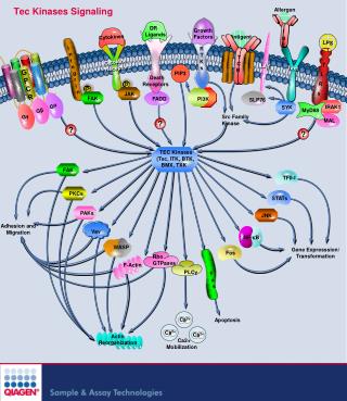
Signaling Cascades • The figure shows the involvement of kinases in cell proliferation and survival. • In this cascade the phosphorylation of each kinase by its upstream kinase serves as a signal for downstream activity. • Inhibiting the pathway through inhibition of kinase involved in the pathway is an attractive proposition Current Medicinal Chemistry, 2008 Vol. 15, No. 29 3037 AURIGENE……Acccelerating Discovery
Imatinib Dasatinib Promise of Kinase Inhibitors Druggable Genome Some Advanced Kinase Inhibitors • Kinases are an attractive target class • Druggability • Early successes (FDA approval of some of the kinase inhibitors) • Possibility of structure guided design • Large number of crystal structures in complex with inhibitors are available Kinome AURIGENE……Acccelerating Discovery
General Structure of Kinases • Bi-lobial structure • N-termial lobe • Mainly made of beta-sheets and connecting loops • One functionally important helix • Both lobes joined by a loop called hinge. • ATP binding pocket is in the interface between the lobes • C-terminal lobe • Mainly made of α-helices • Activation loop spans both N- and C-terminal lobes N-terminal lobe C-terminal lobe AURIGENE……Acccelerating Discovery
…GxGxxG… Helix-C Hinge ATP DFG……APE loop Important Structural Elements • Glycine rich loop • Closes in on the ATP • Helix C • Plays an important role in catalysis • Hinge • Adenosine moiety of the ATP makes bidentate H-bond with this region • Activation loop • Starts with conserved sequence DFG and ends with APE. AURIGENE……Acccelerating Discovery
Orientation of the DFG motif critical for the phosphorylation Hinge Metal Metal γ-phosphate coordinates with the metal Substrate • Activation loop (DFG……APE) provides docking site for the substrate • Highly disordered and usually unresolved in the x-ray structures Phosphate Binding of ATP and Catalysis H-bonds S T Y AURIGENE……Acccelerating Discovery
Close up of the catalytic machinery N-terminal lobe Lys Helix-C ATP Glu Water Asp Metal C-terminal lobe Important Residues • In the active conformation of the kinases, a conserved Lys residue makes a salt bridge with a conserved Glu residue in the middle of the helix-C. • This interaction ensures the positioning of the amino acid Asp (of the DFG motif) to coordinate with the γ-phosphate, the divalent metal ion and catalytic water molecule to facilitate catalysis Salt bridge AURIGENE……Acccelerating Discovery
ATP ATP Inhibitor Inhibitor Kinase Inhibitors • In most cases, inhibitors compete with ATP in order to inhibit the kinase • Such inhibitors are ATP mimetics in the sense that they make interactions similar to what ATP makes. G-loop Hinge Phosphate pocket Ribose pocket AURIGENE……Acccelerating Discovery
Various Subsites in Kinases Schematic of the binding pockets An example of a kinase inhibitor bound in the ATP pocket is shown. Apart from hinge region interaction and solvent interaction, the inhibitor occupies a deeper hydrophobic cavity, also known as selectivity pocket Size of an amino acid preceding the hinge region controls the accessibility to the deeper pocket – Gatekeeper, (Typically Met/Leu/Thr/Ile/Tyr) AURIGENE……Acccelerating Discovery
1nM 10nM 100nM 1μM 10μM Type I Inhibitor- Dasatinib • Dasatinib was developed as a c-Src/BCR-Abl inhibitor but was found to hit many other kinases. • Cross reactivity mainly within the TK family; Approved by FDA Deeper pocket Hinge Solvent Ref: Karaman et. al., NATURE BIOTECHNOLOGY VOLUME 26 NUMBER 1 JANUARY 2008 AURIGENE……Acccelerating Discovery
Helix-C Gly rich loop DFG-Out DFG-In DFG-IN vs DFG-OUT • The activation loop (DFG….APE) has to be IN when the kinase is active – DFG “in” conformation • The DFG loop has been shown to be in an “out” position when kinases are inactive. • This can be exploited in the design of inhibitors. AURIGENE……Acccelerating Discovery
ATP Gleevec DFG OUT DFG IN DFG-IN vs DFG-OUT • Differences between DFG IN and DFG OUT structures are exemplified. • DFG loop in OUT position will clash with phosphate of ATP • When DFG moves to OUT helix-C also moves away creating the pocket shown by bold red arrow. • Gleevec binds to the DFG-OUT conformation of the C-Abl kinase. Helix-C PDB:1T46 AURIGENE……Acccelerating Discovery
Example of Type-II Inhbition Hinge Phe-out conformation Schematic of the binding pockets PDB:1KV1 BIRB-796 binds to p-38 in the Phe-out conformation • The doublet of H-bonds with E-111 (helix-C) and D-207 (DFG loop) backbone is very important • Hence a urea or amide is the common feature in these inhibitors Ref: Karaman et. al., NATURE BIOTECHNOLOGY VOLUME 26 NUMBER 1 JANUARY 2008 AURIGENE……Acccelerating Discovery
Some Known DFG OUT Inhibitors 2ofv Lck – DFG out 2og8 Lck – DFG out 2oo8 Tie – DFG out Bioorg.Med.Chem.Lett. 17: 2886-2889 J.Med.Chem. 50: 611-626 Bioorg.Med.Chem.Lett. 17: 2886-2889 J.Med.Chem. 50: 611-626 2p4i Tie – DFG out 2osc Tie – DFG out 2p2i KDR – DFG out Apart from a hinge binding group, the common feature in these molecules is existence of the bi-aryl amide/urea group which makes interaction with Glu (helix-C) and Asp (DFG loop) AURIGENE……Acccelerating Discovery
Allosteric Kinase Inhibition – Type III • Certain kinases have an allosteric pocket in which an inhibitor can co-bind with ATP • The phosphorylation of the substrate is prevented by unavailability of the catalytic Asp • There are no hinge region interactions in these inhibitors. Helix-C ATP DFG loop AURIGENE……Acccelerating Discovery
A Still Different Type of Inhibitor? • Recently Merck published the co-crystal structure of CHK1 kinase with an inhibitor that is bounds far away from the active site. • DFG loop is has IN conformation, but the inhibitor probably occupies substrate binding site. • Such inhibitors are not being designed yet. They could be results of HTS campaigns. PDB:3F9N AURIGENE……Acccelerating Discovery
SBDD at Aurigene All the structural biology efforts are to aid in more focused medicinal chemistry AURIGENE……Acccelerating Discovery