Download
1 / 12
130 likes | 313 Vues
Investigation of chlorouine-induced keratopathy on in vivo confocal microscopy. Jianjiang Xu, Wenqing Zhu, Jiaxu Hong Eye & ENT Hospital Fudan University Shanghai, China. Title: Investigation of chlorouine-induced keratopathy on in vivo confocal microscopy. Paper ID : 365 Disclosure Block :

E N D
Investigation of chlorouine-induced keratopathy on in vivo confocal microscopy Jianjiang Xu, Wenqing Zhu, Jiaxu Hong Eye & ENT Hospital Fudan University Shanghai, China
Title:Investigation of chlorouine-induced keratopathy on in vivo confocal microscopy Paper ID: 365 Disclosure Block: None of commercial relationships with authors.
Outlines Eye & ENT Hospital Background Objective Methods Results Conclusion
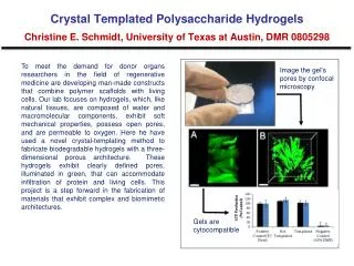
Background • Chloroquine (CHQ) was used successfully for the treatment of several chronic autoimmune diseases • However, a variety of side effects have been reported, of which ophthalmic toxicity is of the utmost importance. Corneal Deposits Chloroquine
Background • In vivo confocal microscopy(CM) has dramatically changed the ophthalmic clinical landscape, allowing a non-invasive approach to in vivo visualize cornea at a magnification of up to ×700 50 µm Cornea Epithelium Stroma Endothelium
To investigate morphological changes of the cornea by CM in patients diagnosed as rheumatoid arthritis and treated with CHQ Objective
Subjects 30 patients with rheumatoid arthritis were enrolled 20 were treated with CHQ the rest served as control Examination: Bilateral eyes were examined by slitlamp biomicroscope and CM CHQ deposits and corneal cellular densities were analyzed Methods
Deposits No deposit was observed by SLB examination. Under CM, CHQ deposits were observed in 28 (70.0%) of 40 eyes in patients CM deposits demonstrate different characteristic features of the CHQ between two groups. Results
Cellular density The cellular density of each layer had no significant difference between two groups except the density of anterior stromal keratocytes. Results
Conclusion • CM is a useful tool for the investigation of keratopathy induced by CHQ treatment • CHQ deposits were mainly identified in corneal epithelium and stroma • They might correspond to intracellular lysosome-like corpuscles which could not be metabolised • Abnormal microenvironment on basal epithelial cells leads to a vicious cycle on the maturation of epithelial cells.
Acknowledgement Team Prof. Jianjiang Xu Wenqing Zhu M.D. Jiaxu Hong M.D. Anji Wei M.D. Gang Li Ph.D Grants: This work was supported partly by grants from the Key Clinic Medicine Research Program, the Ministry of Health, China (2007-2009).