Download
1 / 37
730 likes | 1.45k Vues
CDT A3 lecture. Biomedical Signal Processing An introduction. Prof. Lionel Tarassenko Institute of Biomedical Engineering University of Oxford. Overview of lecture. Types of biomedical signals Biopotentials Probing the body with energy Pre-processing Frequency-based representations

E N D
CDT A3 lecture Biomedical Signal Processing An introduction Prof. Lionel Tarassenko Institute of Biomedical Engineering University of Oxford
Overview of lecture • Types of biomedical signals • Biopotentials • Probing the body with energy • Pre-processing • Frequency-based representations • Feature extraction
Overview of lecture • Types of medical signals • Biopotentials
Biopotential: the electrocardiogram (ECG) • If two surface electrodes are attached to the upper body (thorax), the following electrical signal is observed:
Components of the ECG waveform • P-wave: a small low-voltage deflection caused by the depolarisation of the atria prior to atrial contraction. • QRS complex: the largest-amplitude portion of the ECG, caused by currents generated when the ventricles depolarise prior to their contraction. • T-wave: ventricular repolarisation. • P-Q interval: the time interval between the beginning of the P wave and the beginning of the QRS complex. • Q-T interval: characterises ventricular repolarisation.
Biopotential: the EEG • The electrical activity of the brain (the electroencephalogram or EEG) can be monitored with a pair of scalp electrodes. Its frequency content decreases with alertness. Awake Light sleep Deep sleep
Overview of lecture • Types of medical signals • Biopotentials • Probing the body with energy
Electrical impedance pneumography(measurement of respiration rate) • 4-electrode method to measure electrical impedance (Z) at 20 kHz • Most of the changes in Z across the chest are due to breathing (although some are also caused by blood flow in and out of the heart)
Electrical impedance pneumography • There are no other clinically acceptable methods of monitoring respiration non-invasively • 2-electrode configuration (using ECG electrodes) is sufficient to obtain respiration rate
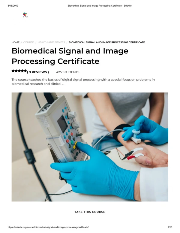
Pulse oximetry(measurement of oxygen saturation) • Oximetry is a technique for measuring how much oxygen the blood is carrying, the oxygen saturation of the blood • The haemoglobin molecule (which is bound to the red blood cells) carries the oxygen in the blood • The two forms of the molecule (Hb and HbO2) have different optical spectra
Pulse oximetry(measurement of oxygen saturation) • The wavelength range between 600 and 1,000 nm is also the range for which there is the least attenuation of light by body tissues. • By measuring the light transmission through a body segment at two wavelengths within that range, the arterial SaO2 can be determined. Oximetry is a non-invasive optical technique
The rationale for pulse oximetry • It is hard to differentiate between absorption due to arterial blood and the absorption due to venous blood, skin tissue and bone. • Two previous solutions: • Compression to obtain a bloodless measurement (not acceptable) • Complex model and even more complex instrument • The solution: pulse oximetry
Pulse oximetry(measurement of oxygen saturation) • Discovered in Japan in the mid-1970's (Aoyagi, 1974) • Only that part of the signal directly related to the inflow of arterial blood into the body segment is used for the calculation of SaO2. • It is assumed that the increase in attenuation of light is caused by the inflow of arterial blood only.
Pulse oximetry(measurement of oxygen saturation) • Light absorption across the finger (or the earlobe) is measured using a single probe with two LEDs and a photodiode
The principles of pulse oximetry • Pulse oximetry assumes that the attenuation of light by the finger can be split into 3 independent components: arterial blood, venous blood and tissues • The amplitudes of the pulsatile component of light attenuation at the two wavelengths are used to derive arterial oxygen saturation
Measurement of blood pressure • Non-invasive blood pressure measurement requires an inflatable cuff and a pressure transducer
Measurement of blood pressure • Blood Pressure is generally described by two measurements (in mmHg): • Systolic Pressure • Diastolic Pressure • Generally quoted as "Systolic over Diastolic". • e.g. 120/70 is a systolic pressure of 120 mmHg and a diastolic pressure of 70 mmHg.
Measurement of blood pressure • Systolic pressure is the arterial pressure when the heart is beating (i.e. during systole). • It is, broadly speaking, the highest pressure present in the arterial (and vascular) system and is a reflection of how hard the heart is pumping. • Diastolic pressure is the arterial pressure when the heart is not beating (i.e. during diastole). • It is, broadly speaking, the lowest pressure present in the arterial system and is a reflection of the resistance against which the heart is pumping.
Measurement of blood pressure • There are two other measures of blood pressure: • Pulse Pressure, the difference between systolic and diastolic pressures. • Mean Arterial Pressure, the mean over the cardiac cycle. It is usually calculated using the following formula: MAP = 2/3 Diastolic Pressure + 1/3 Systolic Pressure
Measurement of blood pressureusing oscillometry • Pulsatile oscillations in cuff pressure are extracted by high-pass filtering of the pressure transducer signal • Max oscillations occur when the cuff pressure is equal to the mean arterial pressure • Systolic and diastolic pressures are identified as occurring at fixed percentages of maximum oscillations (0.55 and 0.85, respectively)
Overview of lecture • Types of medical signals • Biopotentials • Probing the body with energy • Pre-processing
Pre-processing • Medical signals are often corrupted by: • Movement artefact • Muscle noise • Mains frequency noise
Movement artefact • Computation of heart rate is often corrupted by movement artefact • Beat-to-beat heart rate is the reciprocal of the interval between two successive R-peaks in the ECG (x 60 to give beats per minute)
Pre-processing • Medical signals are often corrupted by: • Movement artefact • Muscle noise • Mains frequency noise • Noise can be removed using standard digital filtering techniques; an alternative is the use of Principal Components Analysis to smooth the signal
PCA smoothing • Project data onto the principal component “axes”. • Reconstruct a smoothed signal using only the first few principal axes. • This produces a smooth signal – the remaining axes are mostly noise.
PCA smoothing of ECG waveform • Low numbered axes capture broad shapes • Higher numbered axes give details. • Highest axes correspond mostly to noise
PCA smoothing of ECG T-wave • 2-component reconstruction fails to capture the entire shape • 7-component reconstruction gives smooth and reasonably accurate representation
Overview of lecture • Types of medical signals • Biopotentials • Probing the body with energy • Pre-processing • Frequency-based representations
Short-term Fourier transform • First extract N samples of signal and then window (using Hamming, Kaiser or Hanning windows) to avoid sharp discontinuities at edges. • Then apply the Discrete Fourier Transform [ O(N2) operations ] or the Fast Fourier Transform [ O(N log N) operations] if N is a power of 2.
AR models for spectral estimation • The notation AR(p) refers to the autoregressive model of order p. The AR(p) model is written as follows: X t = ai X t-i + t(1 i p) where the ai’s are the parameters of the model and εt is a white-noise process with zero mean. • An autoregressive model is essentially an infinite impulse response filter which shapes the white-noise input. The poles are the resonances of the filter and correspond to the spectral peaks in the signal.
Overview of lecture • Types of medical signals • Biopotentials • Probing the body with energy • Pre-processing • Frequency-based representations • Feature extraction
Feature extraction • Time-based representation: concatenate values of parameters such as heart rate (x1), breathing rate (x2), oxygen saturation (x3), etc… into a feature vector x = {x1, x2, x3, …} • Frequency-based representation: use encoding provided by AR coefficients (lower dimensionality than FFT coefficients) • Time-frequency based representation: use coefficients from Wavelet Transform
Real time monitoring of vital signs Early alert on Patient condition Intervention by care team Data Fusion Interpretation Heart rate Fusion Single representation of patient status Respiratory rate Oxygen saturation Blood Pressure Temperature Next term: Information-driven healthcare Early identification of physiological deterioration in hospital patients using vital sign data fusion