Download
1 / 47
470 likes | 473 Vues
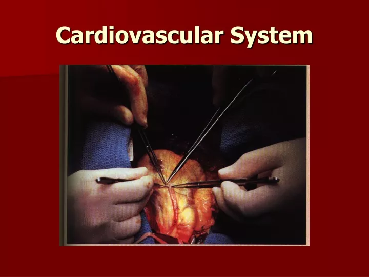
This article discusses the functions of the heart, its general characteristics, coverings, walls, chambers, valves, blood flow, blood supply, and heart physiology.

E N D
I. Functions of the heart. 1. Generating _______________. 2. Routing ________________. 3. Ensuring blood moves __________. 4. _______________ blood supply.
Cardiovascular System II. Heart A. General Characteristics • 1. ____________time/day • 2. ____/day = ____ gallons • 3. ________ miles of blood vessels • 4. Center of the circulatory System • 5. Center of the Thoracic Cavity, between the lungs • 6. ____ on the left side • 7. ______ in size after the age of 65.
B. Coverings of the heart 1. ___________ - covers the heart, 2 main layers a. ___________________(pericardial sac) •Outer •Loose fitting 1. _________ - Outer, thick, tough dense connective tissue. Protects & anchors to the diaphragm 2. _________ - Inner, thin, Squamous epithelium b. _____________________(Epicardium) •Attaches to the surface of the heart •Considered to be the outer most layer of the heart. c. ________________ •Space filled with ________ •_______________ •____________ - swelling of space
Review • What is the main function of the heart? • What is the general size & shape of the heart? • Which cavities contain the heart?
C. Heart Walls ______ layers - middle is the most important - ______________________ • _________ - Thin protective barrier of the heart Serous membrane Fat deposits 2. ___________ -Bulk of the heart _______________- bundles of connective tissue Layer that contracts 3. ___________- Inner most layer Smooth white layer • __________________
D. Heart Chambers Superior • ______________ - Receiving chambers for blood __________role in pumping _________- ridges of muscle _________- separates the atrium into right & left halves _________- oval depression of the heart believed to be an opening in the fetal heart a. ____________- receives from the vena cava b. _____________receives from the lungs
Inferior 2. _______________ - forces blood through the body ____________- Irregular folds of muscle in the endocardium __________________- Slender projections off of the trabuclaecarnae • Attaches to valves & aids in function __________________- Separates ventricles into left & right halves __________________- Externally separates the atrium & ventricles
Review • What are the 3 layers of the heart from outer to inner most? • Why is the inside of the heart coated with simple squamous? • What are the chambers of the heart? • What wall divides the right and left sides of the heart?
E. Heart valves Blood flows in __________________ Prevents ___________________ • 1. _________________(AV) - between atrium & ventricles _______triangular flaps or cusps Point downward into the ventricles ___________ between right Atrium & ventricle (3 flaps) Bicuspid (mitral) between left atrium & ventricle (2 flaps) Chordae tendinae - strands of connective tissue Anchors cusps to papillary walls of the ventricles Murmur - Cusps do not lose completely - leaking of blood
2. Semilunar (SL) Valves Between ventricles & blood leaving Pulmonary & Aortic 3 half moon (semilunar) cusps
G. Supply of blood to the heart Coronary Circulation Right & Left Coronary Arteries - carries fresh oxygenated blood (70% of oxygen, only 25% to skeletal muscles, increases to 70% during exercise) Great & Small cardiac vein Coronary Sinus - large vein that collects blood leaving the heart
II. Heart Physiology - Pumps blood through out the body A. Cardiac Cycle - contraction of both the atria & then ventricle. 1. Systole - Contraction 2. Diastole - Relaxation B. Heart Sounds - LUB - DUB Closing of the heart valves 1. Lub - Closing of the AV valves 2. Dub - Closing of the Sl valves
C. Heart Conduction - Each cardiac cycle is stimulated by special conducting cells in the heart. 1. Receives a signal form the autonomic nervous system. 2. Sinoatrial (SA) Node “Pacemaker” - cluster of pace setting cells Initiates each cardiac cycle by generating an electric impulse. Spread quickly through out the atrium. Stimulates the second cluster of cells.
3. Atrioventricular (AV) Node, AV. bundle, Bundle of His - relays the signal to the ventricles. Extends down the septum of the heart. 4. Purkinje fibers - branches of the AV node, passes further into the myocardium. 5. If the SA node is unable to produce the electrical impulse for the heart to contract, the AV node functions as the pacemaker 6. Slower – Ectopic beat.
D. Electrocardiogram (ECG or EKG) - measures the electrical events during a cardiac cycle 1. Detect changes in the electrical changes in the heart wall 2. Electrical changes produces a changes in the ionic flow through out the body SA node fires send action potential. P Wave - depolarization of the atria - action potential. QRS Wave - depolarization of the ventricle. T Wave ventricular repolarization of the ventricle
PQ interval – atria contract & begins to relax. QT interval – ventricle depolarizes & repolarizes.
E. Cardiac Output - Volume of blood pumped 1. Heart rate X Stroke Volume = Cardiac Output 75 bpm X 70 ml = 5250 ml/min (5.25 L/min) 2. Adjustments - exercise Heart rate Stroke Volume 3. Starling’s Law - Further the heart is stretched, the stronger the contraction. Preload – pressure on the heart when the ventricles are stretched when filling with blood. Afterload – pressure the heart must beat against.
F. Regulation of Heart Activity 1. Controlled by the reflex center (cardioregulatory center) - medulla oblongata. 2. Baroreceptors - detect the blood pressure. 3. Parasympathetic fibers from the medulla oblongata through the Vagus nerve extends to the heart. Acetylcholine (Ach) slows the heart Norepinephrine (NE) speed the heart up
G. Cardiac cycle Three major events in the cycle. 1. Systole – a. Blood is pushed towards the atria, closely the AV valves. b. Pressure increases in the ventricle forcing the SL valves to open. 2. Diastole – a. Pressure in the ventricles decrease, the AV valves open and blood fills the ventricles up to 70% of their volume. 3. End- Atria relaxes & fills with blood, then contract & starts it over.
III. Aging A. By 70 output is reduced by 30%, by 85 30%-60%. B. Hypertrophy is common (enlargement of left ventricle), due to increase afterload (high blood pressure). Leads to decreased elasticity & increased stiffness. Increased left atria pressure and cause pulmonary edema, feel out of breath. C. Greater amount of time to contract & relax leading to decreased in maxmium heart rate. D. Connective tissue with the valves becomes less flexible. E. Development of coronary artery disease in 10% of people over 80.
VI. Cardiovascular diseases • Congestive heart failure - failure of the heart to pump blood to the body tissues. B. Heart Block – Failure of the SA or AV. to generate impulses. C. Heart fibrillation - Heart beats at a irregular pace. D. Heart flutter - heart race up to 300 bpm. E. Hypertension - elevated blood pressure. F. Murmur - Leaking of blood through a closed valve. G. Myocarditis - infection of the heart muscle. H. Pericarditis - Infection of the pericardial sac which results in thicken or scarring.
Blood Vessel and Circulation Functions 1. Carry Blood 2. Exchange of nutrients 3. Transport 4. Regulate blood pressure 5. Direct blood flow
I Blood Vessels - Forms a closed circulatory system (Arteries ->Capillaries ->Veins) Made up of three layers a. Lumen - space for the flow of blood. b. Tunica Intima - inner lining. c. Tunica Media - smooth muscle, contractibility. d. Tunica Adventitia – anchoring
Review • 1. State the functions of blood vessels. • 2. What is the inner space called of blood vessels? • 3. What are the three layers of blood vessels?
A. Arteries - Carries blood away from the heart Strong & Elastic 1. Elastic Arteries – Largest in diameter. Mainly elastic 2. Muscular arteries – medium sized and small diameter. Mainly smooth muscle 3. Distributing arteries – vasoconstriction - Vasomotor fibers of the (autonomic). vasodilatation - nerve impulse is inhibited - muscle relaxes & elastic fibers recoils. 4. Small arteries 5. Arterioles - 0.5 mm in diameter
Review • 1. What are the function of arteries? • 2. Why are arteries thick? • 3. How are arteries adapted to handle high blood pressure?
B. Capillaries - Thin wall blood vessels that permit exchanges of material. 1. Connect arteries to veins 2. 0.01 mm in diameter - lumen 3. Can only fit 1 RBC at a time. 4. Form capillary beds or networks. • Thoroughfare channels - connect arterioles directly to veins • True Capillaries - 10-100 per bed Precapillary Sphincter - valve that regulates flow of blood into those capillaries.
Review • 1. What is the function of capillaries? • 2. How are capillaries adapted for their function? • 3. Describe a Thoroughfare channels.
C. Veins - Carries blood toward the heart 1. Thinner & collapse. 2. Holds 65% of the body’s blood. 3. Low blood pressure, too low to be pumped back to the heart. 1 way valves Body movement - Skeletal contraction, breathing. 4. Made up of three layers - same as arteries 5. Varicose veins - overstretched veins from blood pooling , hemorrhoids. 6. Venules – thinner versions of veins
Review • What is the function of veins? • How are veins adapted to handle low pressure? • Name some ways that blood can be “PUSHED” through the veins.
II. Blood Pressure - force exerted by blood against the inner walls of vessels A. Influenced by: Cardiac Output Blood Volume Peripheral Resistance which is regulated by nerve, kidneys, hormones. B. Moves from regions of higher pressure to lower pressure Systolic - Peak pressure - 120 mmHg Diastolic - Resting - 70 - 80 mmHg Sphygmomanometer - measures blood pressure. Stethoscope – Hearing Korotkoff - tapping sound sounds
C. Pulse - rhythmic expanding & recoiling of an arterial wall Average Adult 70 - 90 bpm Child 80 - 140 bpm tachycardia - heart rate is above 100 bpm at rest bradycardia - heart rate is lower than 60 bpm at rest Auscultatory Method Sphygmomanometer Stethoscope Korotkoff sounds
Review • Define blood pressure. • Describe blood pressure movement. • Define pulse.
III Capillary Exchange 10 Billion capillaries Materials (gases, nutrients) move across capillary walls by diffusion, osmosis, facilitated diffusion & active transport. Small amount of fluid also moves across the capillary walls: Hydrostatic pressure - blood pressure within the capillaries Osmotic pressure - movement of fluid from cells to plasma 90% reabsorbed back into the blood 10% returned back by the lymphatic system
Review • What are the functions of capillaries? • What is the purpose of hydrostatic pressure? • How does hydrostatic pressure compare to osmotic pressure? • Which part of the capillary bed has the highest hydrostatic pressure and osmotic pressure?
IV. Regulation of blood pressure - regulation of steady pressure is important. A. Nervous Control - Adjusts cardiac output & peripheral resistant by autonomic fibers to the SA node & reflex center (vasomotor center in the medulla oblongata & pons). 1. Peripheral resistance - control by activity of vasomotor between the smooth muscle & reflex center of Medulla (Vasomotor tone, increases = constriction). Control of vasomotor center (MAP) Map = CO X PR Baroreceptors – located in major vessels above the heart & detect changes in the blood pressure. Moment to moment control
b. Chemoreceptors - Sensitive to changes in oxygen levels or hydrogen ions (pH) level. carotid bodies – carotid sinus aorta bodies - aorta
Review • What is the job of baroreceptors & chemoreceptors?
V. Cardiovascular diseases Aneurysm - formation of a sac within the heart or blood vessels due to stretching B. Arteriosclerosis - Loss of elasticity in arterial walls C. Atherosclerosis - narrow of arteries by plaque build up
1. What are the three types of blood vessels and how are they different? 2. What is the difference between vasodialation & vasoconstriction? 3. What are the 3 layers of the blood vessels? 4. Does blood ever flow in reverse within the blood vessels?