Download
1 / 9
90 likes | 182 Vues
The Digestive System … Notes II. The Digestive Process. …see “Notes” handout.

E N D
The Digestive System … Notes II The Digestive Process …see “Notes” handout
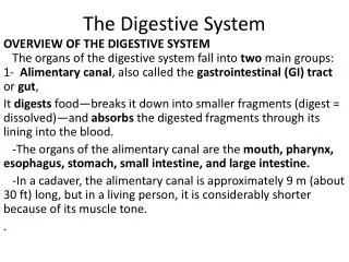
When food is ingested, it is put into the mouth where it is chewed. Chewing is a physical digestion process that increases the surface area so that chemical digestion can take place faster. Connected to the mouth are a series of salivary glands, which release saliva through ducts (tiny tubes) that lead into the mouth. Saliva contains water and an enzyme. The water acts both as a lubricant to aid in swallowing and as a reactant in the hydrolytic reactions of digestion. The enzyme, salivary amylase (ptyalin), breaks the ester bonds between the sugar molecules of starch and begins the chemical conversion of cooked starch into maltose. The tongue is a muscle that rolls the food into swallow able sized balls, each one called a bolus, and pushes them to the back of the mouth from where swallowing takes place.
The chamber at the back of the mouth is called the pharynx. It is a common chamber for both food and air. The trachea and the esophagus split off from the base of the pharynx and transport their contents to their respective destinations. In order to prevent the food materials from going down the trachea, the epiglottis, a ventral flap of tissue, covers it. The muscle contractions moving the bolus are called peristalsis. At the base of the esophagus, the bolus encounters a constriction, the cardiac sphincter, that must relax and open before the bolus can enter the stomach. The stomach is a large "J-shaped" organ. Its walls have three layers of muscle that churn the food materials over and over. The presence of food in the stomach causes the release of gastrin, a hormone, which travels from the walls of the stomach into the blood stream.
As gastrin circulates the body, it affects the stomach and causes the release of gastric juice. Gastric juice contains water as well as HCI and pepsinogen. Under the influence of these, the bolus becomes acid chyme (chyme, literally, means "runny"). The HCI has two functions. It creates an environment with a low pH (about 2.5) that will kill any bacterial growth that may be on the food material. HCI also reacts with pepsinogen and converts it into pepsin, a protease. Pepsin is secreted as the precursor pepsinogen rather than in its final active form. Being a protein digesting enzyme, a cell would not survive the production of active pepsin. The stomach walls produce mucous to protect themselves from the pepsin. Deterioration of the mucous lining of the stomach results in an ulcer.
The pyloric sphincter is similar to the cardiae sphincter, except it is located at the base of the stomach where it controls the passage of the liquid acid chyme, a small amount at a time, into the duodenum (first part of the small intestine). The duodenum is specialized by the presence of chemoreceptors, chemical sensitive nerve endings that are able to detect the different biochemicals in the food material. In this way, the digestive system can regulate which secretions are released. The presence of the acid chyme triggers the release of secretin from the duodenum. Secretin is a hormone that travels through the blood to the pancreas where it causes the release of pancreaticjuice. Sodium bicarbonate is a component of pancreatic juice. Bicarbonate ions (HCO31-) over neutralize the acid chyme and it becomes alkaline with a pH of about 8.5. The other components of pancreatic juice are enzymes that are active at this alkaline pH.
The presence of lipids in acid chyme causes the release of a hormone called CCK (cholecystokinin). CCK affects both the gall bladder and the pancreas. It causes the release of bile and pancreatic juice containing the lipase enzymes that will digest lipids. • The enzymes in pancreatic juice are: • lipase - converts lipids into fatty acids and glycerol • trypsin - a protease that breaks many peptide bonds to convert protein into peptides of varying lengths • pancreatic amylase which breaks remaining starches into maltose molecules • nucleases - which partially convert nucleic acids into nucleotides
These enzymes are active in the duodenum. The small intestine also produces and secretes its own digestive enzymes. The hydrolysis of disaccharides by enzymes like maltase completes the digestion of carbohydrates. Peptidases break any remaining peptide bonds in the proteins to release amino acids, the unit molecules. The last part of the small intestine is called the ileum. The ileum is specialized to absorb the products of digestion. It has a huge surface area, which maximizes the potential for absorption. It is also lined with specialized structures for absorption. These structures are called villi
The products of fat digestion enter the lacteals, part of the lymphatic system. The rest of the products of digestion enter the blood stream. The cells lining the villi are equipped with lots of mitochondria to produce the A TP energy required for the active transport of these products of digestion. Once the nutrient value from the food is absorbed, the remains are indigestible materials (cellulose), water, and other components of the food that the body didn't require. These things pass from the ileum through the ileo-caecal valve (another sphincter) into the large intestine ( colon). The first segment of the colon is called the caecum. Extending down from the caecum is the appendix.
The major roles of the colon are to absorb a great amount of the water that was added to the food material all the way along the digestive system, and to house E. coli. E. coli is a bacterium that metabolizes some of what our bodies were unable to. It lives in symbiosis with humans because it is able to obtain its nutrients from the waste material. Part of what it does is release minerals, manufacture vitamins and amino acids, which get absorbed along with the water into the circulatory system. The bacteria begin the decomposition of the waste materials and convert them to feces with a changed color, smell, and texture. The last part of the colon is the rectum that stores feces until defecation. It ends with the anal sphincter, which controls defecation.