Download
1 / 34
430 likes | 2.17k Vues
DENTAL ANATOMY. BY DR. MANISHA MISHRA. Oral Cavity Entrance of the gastrointestinal tract Dental arch: alveolar arch of the maxilla and mandible 4 types of teeth Incissor Canine Premolars Molars. Boundaries. Anterior: Lips

E N D
DENTAL ANATOMY BY DR. MANISHA MISHRA
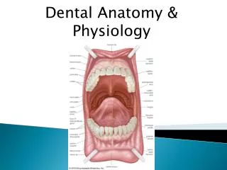
Oral Cavity • Entrance of the gastrointestinal tract • Dental arch: alveolar arch of the maxilla and mandible • 4 types of teeth • Incissor • Canine • Premolars • Molars
Boundaries • Anterior: Lips • Posterior: anterior tonsillar pillars (palatoglossal folds) • Lateral: cheeks • Roof: palate • Floor: anterior 2/3 of tongue
Parts • Oral vestibule • Space between the lips and teeth/gingiva • Oral cavity proper -
Functions • Speech • Respiration • Digestion • Chewing • Aesthetic
Landmarks • Frenulum • Mucobuccal fold (posterior) • Mucolabial fold( ant ) • Canine eminence • Parotid papilla ( stenson’s duct ) • External oblique ridge • Retromolartrigone
Landmarks on roof • Incisive papilla: covers the incisive foramen between 2 incissors • Palatine rugae • Uvula
Landmarks on the floor • Tongue • Lingual sulcus • Frenulum
Anatomy of tooth • Anterior teeth: Incissors and canine • Posterior teeth: Molars and premolars Each tooth consists of: • Crown – The part which protrudes from the gum • Root – Part embedded in the bone • Neck – Slightly narrowed region where the crown merges with root
Components of tooth • Pulp containing neurovascular bundle • Dentine • Enamel • Cementum
enamel • Hardest tissue • Surrounds dentine • highly mineralised Main mineral- hydroxyapatite
dentine • Dentine is the most abundant dental tissue • Determines the size and shape of teeth. • Dentine is a bone-like substance that is formed by odontoblast cell which makes up most of the structure of the tooth. • Dentine is found just under the enamel in the crown and under the cementum in the root..
pulp • Connective tissue, mainly consits of odontoblast and fibroblast • Contains neurovascular components • There is anastomosis between arterioles & venules
cementum • Cementum is a specialized bony substance covering the root of a tooth • Cementum is excreted by cells called cementoblast within the root of the tooth • Its color is yellowish and it is softer than either dentine or enamel. Alveolar Bone • Mineralized tissue that surrounds the teeth in jaws
Periodontal ligament • The periodontal ligament is a specialized connective tissue that attaches a tooth to the jaw bone Function • Helps tooth withstand large compressive forces which occur during chewing, without destruction of the adjacent alveolar bone • to serve as a source of sensation. (outer covering of the tooth (enamel) has no sensory receptors itself.)
gum • Mucous-membrane-covered connective tissue attached to and surrounds the neck and the alveolar bone. • Edges of the gums around the teeth are free and extend into the spaces between the teeth. • healthy gum is pink and tough. • have limited sensitivity to - pain, Temperature, pressure
Blood supply • Periodontal plexus • Inferior/superior alveolar vessel • Periosteal vessel • Vessel from adjacent musculature
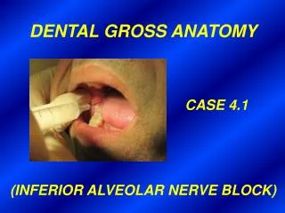
Nerve supply • Upper teeth – maxillary nerve (Ant. Sup. Alveolar, middle sup. Alveolar & post. Sup. Alveolar nerves) • Lower teeth – mandibular nerve (inferior alveolar nerve)
Mandible Only moving bone present in skull, Has sockets for lower teeth • Parts – Body & 2 Rami Body – • 2 surfaces – External & Internal • & 2 borders – Upper (Alveolar), Lower(Base) Ramus • 2 Processes – Coronoid & Condyloid
Mandibular Foramen - opening on the medial surface of the ramus • Oblique Line - located on the superior lateral surface of the body • Mental Foramen - foramen for the transmission of the mental nerve (cutaneous to the lower third of the face) and artery
The mandible is the body support for the mandibular teeth • And is also the insertion for the four primary muscles of mastication and the accessory muscles of mastication. • It is the movable portion of the TMJ articulation with the temporal bone
The Muscles of Mastication - The chief muscles of mastication are: • Masseter. • Medial Pterygoideus • Lateral Pterygoideus • Temporalis. Nerve • Mandibular division of Trigeminal Nerve
maxilla • The maxilla is a fusion of two bones along the palatal fissure that form the upper jaw • has sockets for upper teeth
Parts • Body • Four processes • The zygomatic process • The frontal process • The alveolar process • The palatine process
NOMENCLATURE • DECIDUOUS DENTITION: • Central incisior=A • Lateral incisior=B • Canine=C • 1StDecidious molar=D • 2ndDecidious molar=E
PERMANENT DENTITION: • Central incisior=1 • Lateral incisior=2 • Canine=3 • 1st premolar=4 • 2nd premolar=5 • 1st permanent molar=6 • 2nd permanent molar=7 • 3rd permanent molar=8