1 / 15

Renal Anatomy

Renal Anatomy. Tony Tiemesmann Dept Diagnostic Imaging Bloemfontein Hospital Complex. Contents. Embryology Macroanatomy Microanatomy Imaging anatomy XR Ultrasound CT MRI. Embryology 1. Pronephros Mesonephros Metanephros Pronephric duct from intermediate mesoderm – “ glomeruli ”

yehuda
Télécharger la présentation

Renal Anatomy

An Image/Link below is provided (as is) to download presentation Download Policy: Content on the Website is provided to you AS IS for your information and personal use and may not be sold / licensed / shared on other websites without getting consent from its author. Content is provided to you AS IS for your information and personal use only. Download presentation by click this link. While downloading, if for some reason you are not able to download a presentation, the publisher may have deleted the file from their server. During download, if you can't get a presentation, the file might be deleted by the publisher.

E N D

Presentation Transcript


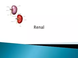
  1. Renal Anatomy Tony Tiemesmann Dept Diagnostic Imaging Bloemfontein Hospital Complex

  2. Contents • Embryology • Macroanatomy • Microanatomy • Imaging anatomy • XR • Ultrasound • CT • MRI

  3. Embryology 1 • Pronephros • Mesonephros • Metanephros • Pronephric duct from intermediate mesoderm – “glomeruli” • Mesonephric duct with ureteric bud – “collecting system”

  4. Embryology 2

  5. Macroanatomy 1

  6. Macroanatomy 2

  7. Macroanatomy 3

  8. Macroanatomy 4

  9. Microanatomy

  10. Imaging: X-rays

  11. Imaging: Ultrasound

  12. Imaging: CT 1

  13. Imaging: CT 2

  14. Imaging: MRI

  15. References • Daly KP et al. Traumatic Retroperitoneal Injuries. RadioGraphics. October 2008 28, 1571-1590 • Learningradiology.com • Radiologyassistant.nl • Ryan S et al. Anatomy for diagnostic imaging 3rd edition. Saunders-Elsevier. 2011

More Related