Download
1 / 44
440 likes | 444 Vues
CHAPTER 40 AN INTRODUCTION TO ANIMAL STRUCTURE AND FUNCTION. Introduction Hierarchy of Organization Tissue Types Organ Systems Homeostatic control. The study of animal form and function is integrated by the common set of problems that all animals must solve these are the needs

E N D
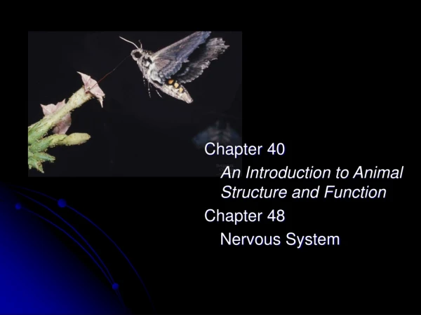
CHAPTER 40AN INTRODUCTION TO ANIMAL STRUCTURE AND FUNCTION Introduction Hierarchy of Organization Tissue Types Organ Systems Homeostatic control
The study of animal form and function is integrated by the common set of problems that all animals must solve these are the needs Gas Exchange Providing Nourishment Excreting waste reproduction Form vs. Function
Remember; Form/Function branches of biology; Anatomy is the study of the structure of an organism. Physiology is the study of the functions an organism performs.
We have studied prokaryotes – the simplest cells. Now we are looking at some of the most complex cells. Larger, many organelles and compartments. Why Specialize??
Who would have the toughest time in life: A “normal” sized adipose cell A severely engorged (enlarged) adipose cell WHY??
For weekend Read about “HPA axis” What is its function? How is its function regulated (mechanisms)?
specialized cells perform microscopic tasks. Tissues are groups of cell with a common structure and function Tissues combine to perform specific macroscopic tasks organs. Organ systems organismecosystemplanetplanetarysystemgalaxygalaxyclusteruniversemultiverse Studying Form:hierarchical levels of organization
A - Epithelial Tissue B - Connective Tissue C - Nervous Tissue D - Muscle Tissue Our goal identify and characterize cells for each tissue III - Primary Tissue Types of all animals
A - Epithelial Tissue (surface tissue) is classified by the number of cell layers and the shape of the cells on the free surface. Types of layers; A simple epithelium has a single layer of cells, and a stratified epitheliumhas multiple tiers of cells. Shapes of cells; cuboidal (like dice), columnar (like bricks on end), or squamous (flat like floor tiles).
Depending on Surface, Epithelial cells HIGHLY Specialized EXAMPLES: Glandular epithelia lining tubules in the thyroid gland secrete a hormone that regulates fuel consumption (thyroxine). Glandular epithelia that line the lumen of the digestive and respiratory tracts secretes mucus, lubricating the surface and keeps it moist. The free epithelial surfaces of some mucous membranes have beating cilia that move the film of mucus along the surface. In the respiratory tubes, this traps dust and particles.
B - Connective tissue functions mainly to bind and support other tissues. Connective tissues have a sparse population of cells scattered through an extracellular matrix. The matrix generally consists of a web of fibers forming in a uniform foundation that may be liquid, jellylike, or solid. Cells are held in this matrix In most cases, the connective tissue cells secrete the matrix.
There are three kinds of connective tissue fibers (matrix) Collagenous fibers are made of collagen. Collagenous fibers are nonelastic and do not tear easily when pulled lengthwise (strong). Elastic fibers are long threads of elastin. Elastin fiber provide a rubbery quality (flexible). Reticular fibers are very thin and branched. Composed of collagen and continuous with collagenous fibers, they form a tightly woven fabric that joins connective tissue to adjacent tissues (net-like).
The major types of connective tissues loose connective tissue, adipose tissue, fibrous connective tissue, cartilage, bone, and blood. Fig. 40.2
1. Loose connective tissuefunction binds epithelia to underlying tissues and functions as packing materials, holding organs in place (mesentery). Loose connective tissue has all three fiber types. Two cell types predominate in the fibrous mesh of loose connective tissue. Fibroblasts secrete protein matrix Macrophages amoeboid cells - delta force...know about these!!
2. Adipose tissue is a specialized form of loose connective tissues that store fat in adipose cells distributed throughout the matrix. Adipose tissue pads and insulates the body and stores fuel as fat molecules. Each adipose cell contains a large fat droplet that swells when fat is stored and shrinks when the body uses fat as fuel – physiology of adipose cells intense!!! Visceral fat, type II diabetes. Question - is obesity related to too many adipose cells, or larger than normal fat droplets??
3. Fibrous connective tissue is dense, due to its large number of collagenous fibers. The fibers are organized into parallel bundles, an arrangement that maximizes nonelastic strength. This type of connective tissue forms tendons, ligaments cartilage bone.
4. Cartilage - collagenous fibers embedded in a rubbery matrix - chondroitin sulfate, a protein-carbohydrate complex. No vessels, no nerves (nutrient supply??) Chondrocytes (cells) secrete collagen and chondroitin sulfate. The composite of collagenous fibers and chondroitin sulfate makes cartilage a strong yet somewhat flexible support material. The skeleton of a shark is made of cartilage and the embryonic skeletons of many vertebrates are cartilaginous. Who takes chondroitin and glucosamine supplements???? (my dogs, horses and I)
5. Bone, a mineralized connective tissue. Osteoblasts (cells) deposit a matrix of collagen. Then, calcium, magnesium, and phosphate ions combine and harden within the matrix into the mineral hydroxyapatite. The combination of hard mineral and flexible collagen makes bone harder than cartilage without being brittle. The microscopic structure of hard mammalian bones consists of repeating units called osteons. Each osteon has concentric layers of mineralized matrix deposited around a central canal containing blood vessels and nerves that service the bone.
6. Blood!?!? (a connective tissue?) functions differently from other connective tissues, but it does have an extensive extracellular matrix. The matrix is a liquid called plasma, consisting of water, salts, and a variety of dissolved proteins. Suspended in the plasma are erythrocytes (gas transport), leukocytes (immunity) and cell fragments called platelets (clotting).
In Your Groups Hormones produced/secreted (focus on those influencing HPA) Stimulus to release Primary effects of hormones Briefly brief class As a class Have you heard of cortisone treatments? Long term effects?
C - Nervous tissue senses stimuli and transmits signals from one part of the animal to another. The functional unit of nervous tissue is the neuron, or nerve cell. It consists of a cell body and two or more extensions, called dendrites and axons. Dendrites transmit nerve impulses from their tips toward the rest of the neuron. Axons transmit impulses toward another neuron or toward an effector, such as a muscle cell. Fig. 40.3
D - Muscle tissue composed of long cells called muscle fibers Arranged in parallel within the cytoplasm of muscle fibers are large numbers of myofibrils made of the contractile proteins actin and myosin. Muscle is the most abundant tissue in most animals, and muscle contraction accounts for most of the energy-consuming cellular work in active animals.
There are three types of muscle tissue in the vertebrate body: skeletal muscle, cardiac muscle, and smooth muscle. Fig. 40.4
Attached to bones by tendons, skeletal muscle is responsible for voluntary movements. Skeletal muscle is also called striated muscle. What causes this?:::: Cardiac muscle forms the contractile wall of the heart (myocardium). Striated, branched. The ends of the cells are joined by intercalated disks
Smooth muscle, which lacks striations, is found in the walls of the digestive tract, urinary bladder, arteries, and other internal organs. The cells are spindle-shaped. They contract more slowly than skeletal muscles but can remain contracted longer. Controlled by different kinds of nerves than those controlling skeletal muscles, smooth muscles are responsible for involuntary body activities. These include peristalsis of the digestive system and vasoconstriction.
Synapse Identify differences between neuron/neuron and neuromuscularsynapses. Afferent vs. efferent, motor neurons, peripheral vs. central… Role of neurotransmitters, examples. Sympathetic vs parasympathetic nervous system Receptor mechanisms – all act as terminal end of synapse!! Baro (most are variation of this) Nociceptor Rods/cones Summary points of nervous, muscle tissue
garden slug Arion hortensis Everyone’s favorite Whatever the shiny substance is, the slug produces it. The slug does not have an internal respiratory system, so its “skin” must provide that function. If the slimy slug is sprinkled with salt, it frantically wiggles in seeming pain..and dies. Predict the type of tissue, cells, and structure found on the surface f the slug. EXPLAIN your reasoning. Thank you
In all but the simplest animals (sponges and some cnidarians) different tissues are organized into organs. Many vertebrate organs are suspended by sheets of connective tissues called mesenteries in body cavities moistened or filled with fluid. Mammals have a thoracic cavity housing the lungs and heart that is separated from the lower abdominal cavity by a sheet of muscle called the diaphragm. IV - The organ systems of animals are interdependent
In some organs the tissues are arranged in layers. Vertebrate stomach - four major tissues layers. A thick epithelium lines the lumen and secretes mucus and digestive juices into it. Outside this layer is a zone of connective tissue, surrounded by a thick layer of smooth muscle. Another layer of connective tissue encapsulates the entire stomach. Fig. 40.5
Example of tissue layers Example of tissue layers gone wrong!! This could be any of us now!!
Organ systems carry out the major body functions of most animals. Each organ system consists of several organs and has specific functions.
The efforts of all systems must be coordinated for the animal to survive. Our goal (and the goal of the entire medical community!) is to understand this coordination Assignment Questions for Friday (to be discussed) – record notes in you notebook .. Be prepared to speak! Explain how the nervous system conducts information through both electrical and chemical energy Identify the difference between epithelium and endothelium - provide examples. Pages in the Book: 1025-1035 Words to use in question One • Action/Resting potential • Sodium potassium pump • Threshold potential • Myelin • Schwann cells • Gated channels • Refractory period • Neurotransmitter • Synaptic cleft
The efforts of all systems must be coordinated for the animal to survive. Our goal (and the goal of the entire medical community!) is to understand this coordination Assignment Questions for Wednesday Now that we have an idea of tissue types, where do they come from ( three cell layers during development)? Do all animals develop these tissues in a similar fashion? Can now you predict how spina bifida might occur? What do Marfan’s syndrome, scurvy, and E-bola infections all have in common? Do their symptoms make sense? References – page 639, chapter 47…
Assignment for Thursday Study nerve AP transmission p 1052 Study NM junction events p 1055 Study muscle contraction Study Differences between three muscle types Study Where is Energy Used?????????????????? Can these events be manipulated??? Study
Dendrite Cell Body Axon Neuron
Resting Potential Threshold Depolarization Repolarization Hyperpolarization Undershoot Refractory Action Potential
Na/K pump Na/K gates Stimmuli Mechanical Heat Receptor Voltage Where Important ? Ion ChannelsAnd Pumps
Saltatory conduction White Matter – meylin – where do we see this?? Specialized Conduction
Diagram a neuron. Label three main parts. Show direction of flow For each numbered section, identify direction of ion flow ActionPotential Quiz 3 2 1 4
Synapse: ElectricalChemicalElectrical What happens here? What is a thought…a dream…a memory?
Nicotine/Meth – increased dopamine release/ activate dopamine receptors Cocaine – inhibit acetylcholine esterase LSD Pain Medications – Latacane and other “canes” Alcohol – GABA, hyperpolarization Drugs and Synapse
Assignment Questions Friday (to be discussed-Monday) – record notes in you notebook. Be prepared to speak !!!!!!!!!!!!!!!!!!!!!!!!!!!!!!!!!!!!!!!!!!!!!! 1. Explain the process of muscle contraction (on a cellular level)…what is a rigor state..why does it hurt? 2. Identify specific form function adaptations of the muscle tissues. Neuro-muscular Junction (best from internet) Acetyl Choline (ACH) Calcium T-Tubules Sarcoplasmic Reticulum Troponin Tropomyosin Actin Myosin Smooth vs. Skeletal vs. Cardiac Muscle