Download
1 / 144
1.46k likes | 1.98k Vues
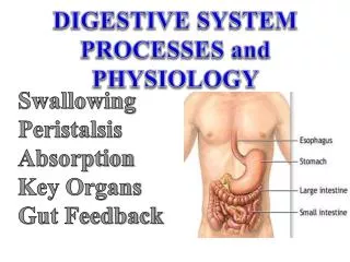
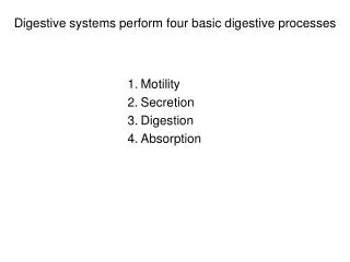
Digestive systems perform four basic digestive processes. Motility Secretion Digestion Absorption. 1. Motility. *Muscular contractions within the gut tube and move forward the contents of the digestive tract *smooth muscle in the digestive tract walls maintain

E N D
Digestive systems perform four basic digestive processes Motility Secretion Digestion Absorption
1. Motility *Muscular contractions within the gut tube and move forward the contents of the digestive tract *smooth muscle in the digestive tract walls maintain a constant low level of contraction: tone *tone is important for: -maintaining a steady pressure on contents -preventing the walls from remaining permanently stretched
Two types of digestive motility Propulsive movements: propel (push) the contents through digestive tract at varying speeds, with the rate of propulsion dependent on the functions of different regions Mixing movements: a. mix food with digestive juices b. facilitate absorption by increasing interactions between contents and surfaces of tract
Contraction sites and control *smooth muscle: most of digestive tract skeletal muscle: ends of tract *acts of chewing, rumination, defecation: motor cortex control (“voluntary”) smooth muscle-contraction: complex autonomic (“involuntary”)
2. Secretion *a number of digestive juices are secreted into the digestive tract lumen by specific exocrine glands, after appropriate neuronal or hormonal stimulation *digestive secretion consists of water, electrolytes, and specific important organic constituents (enzymes, bile salts or mucus) *secretory cells extract materials from blood *secretion requires energy (active transport and synthesis) *digestive secretions are reabsorbed back into blood (may not be in original forms)
3. Digestion *carbohydrates, proteins, and fats are large molecules unable to cross plasma membrane -> need to be broken down into smaller absorbable units by digestive enzymes before entering blood *digestion begins at anterior tracts and is accomplished by enzymatic hydrolysis *digestive enzymes are specific in the bonds they can hydrolyze
stepwise enzymatic breakdown of foodstuffs Table 14-1, p.616
Dietary molecules Carbohydrates monosaccharides polysaccharides starch: enzymes are most widely used cellulose: most abundant organic molecules in biosphere (major part of plant) chitin glycogen
Table sugar Milk sugar Table 14-1a, p.616
Dietary molecules Neutral fats Also, nucleic acids -> pancreatic nucleases -> nucleotides Table 14-1b, p.616
4. Absorption *after digestion, absorption occurs in the middle to posterior sections of tracts *small units of nutrients, water, vitamins, and electrolytes are transferred from lumen into blood or body cavity *specialized transporters in epithelial cells carry hydrophilic units through membrane; surface area enhances absorption (ex. villi)
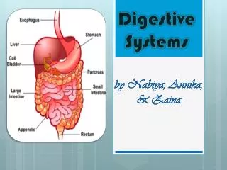
Digestive system: Digestive tract and accessory digestive organs *digestive (gastrointestinal) tract: mouth, pharynx (throat), esophagus, stomach, small intestine, large intestine, anus -> continuous tube but regional differences *accessory organs: salivary glands, exocrine pancreas, the biliary system (liver and gallbladder) -> connected with the digestive tract with ducts
Regulation of digestive function: *digestive motility and secretion are carefully regulated to maximize digestion and absorption Autonomous smooth-muscle function *some smooth-muscle cells are “pacesetter” cells: interstitial cells of Cajalthat display rhythmic, spontaneous variations in membrane potential *slow-wave potential, or basic electrical rhythm (BER), or pacesetter potential *slow-wave oscillations: cyclical variations in Ca2+ release from ER and Ca2+ uptake by mitochondria
Regulation of digestive function: *digestive motility and secretion are carefully regulated to maximize digestion and absorption Autonomous smooth-muscle function *rhythmic cycles of muscle contraction depends on mechanical, nervous, and hormonal factors *gap junctions: transmit electrical activity to contractile smooth muscle cells *rate of contraction: depends on pacesetter cells of each organ
Regulation of digestive function: Intrinsic nerve plexuses: digestive system’s “intramural” nervous sytem (enteric nervous system) a. myenteric plexus b. submucous plexus *located entirely within the digestive tract wall and run its entire length *influence all facets of digestive tract activity -> sensory neurons (input), neurons that innervate muscle or exocrine/endocrine cells (output), interneurons -> acetylcholine vs. NO and vasoactive intestinal peptide
Regulation of digestive function: 3. Extrinsic nerves *nerve fibers from autonomic nervous system that innervate different digestive organs *influence digestive motility and secretion by: regulate intrinsic plexuses, gastrointestinal hormone secretion, smooth muscle and glands directly *“fight-or-flight” sympathetic system vs. “rest-and- digest” parasympathetic system *autonomic nervous system can modify only digestive system -> specific activation of extrinsic innervation leads to coordination of activity between dif. regions
Regulation of digestive function: 4. Receptor activation *three different types sensory receptor: a. chemoreceptors b. mechanoreceptors (pressure receptors) c. osmoreceptors *receptors active through neural reflexes & hormone secretion *digestive system’s effector cells are activated: smooth muscle cells, exocrine gland cells, & endocrine gland cells
Mouth: obtaining and receiving food *mouth, or oral cavity, is ringed by muscular lips -> prehension *palate: hard palate vs. soft palate -> separates mouth from nasal cavity *uvula: sealing off nasal passage during swallowing *tongue: floor of mouth, consists of voluntarily controlled skeletal muscle taste buds (also in soft palate, throat, linings of cheeks) *pharynx (throat): rear of oral cavity -> link between mouth and esophagus -> common passageway for both digestive system and the respiratory system
Mouth: mastication *motility of the mouth that involves slicing, tearing, grinding and mixing food by teeth *teeth: incisors, canines, molar *purposes of mastication: break food, mix food with saliva, stimulate taste buds *taste buds stimulation: reflexly increases salivary, gastric, pancreatic, and bile secretion *food in the mouth -> activates mechanoreceptors -> medullar oblongata has a chewing center -> activates motor neurons that open the oral cavity
Saliva aids in mastication & lubricates food *saliva is secreted by salivary glands outside of mouth *salivary glands: produce serous product (water + enzyme) and mucus *saliva: 99.5% water and 0.5% electrolytes and protein *functions: a. facilitate swallowing: moistening and lubrication b. salivary amylase: starch -> maltose c. antibacterial action: lysozyme and rinsing away bacterial food source d. solvent for molecules that stimulate taste buds e. keeps oral cavity moist f. rich in bicarbonate buffers -> neutralization
Salivary secretion: simple and conditioned reflexes *salivary secretion in vertebrates is the only digestive secretion entirely under neural control (all others also require hormones) *basal secretion: low level, stimulated by parasympathetic (0.5 ml/min, for moisture; max: 5 ml/min) *enhanced stimulation by two different salivary reflexes: 1. Simple (unconditioned) reflex 2. Acquired (conditioned) reflex learned response based on previous experience
+ + + + + Cerebral cortex Other inputs Salivary center in medulla Conditioned reflex extrinsic Pressure receptors and chemoreceptors in mouth Autonomic nerves Simple reflex Salivary glands Salivary secretion Fig. 14-6, p.622
Salivary secretion: simple and conditioned reflexes *both sympathetic and parasympathetic stimulation (autonomic nervous system) increases salivary secretion, but quantity and consistency of saliva changes with food (sensory evaluation of food) *parasympathetic: increases blood flow, prompt and abundant flow of watery saliva rich in enzymes *sympathetic: reduces blood flow -> output of smaller volume but rich in mucus (stress situations) Mouth: digestion is minimal and no absorption
Pharynx, esophagus, and crop *motility associated with the pharynx and esophagus is swallowing or deglutition (food from mouth to stomach) *swallowing begins when bolus is moved into esophagus *pharyngeal pressure receptors -> swallowing center in medulla -> muscles involved in swallowing *swallowing: a sequentially programmed all-or-none reflex highly coordinated activities are initiated in a regular pattern over a period of time
Swallowing: oropharyngeal & esophageal stages *when in pharynx, bolus must be prevented from entering the mouth, nasal passages, and trachea a. tongue against hard palate b. uvula is elevated against the throat, sealing off nasal passages c. elevation of larynx and tight closure of glottis (vocal folds across the laryngeal opening) d. epiglottis e. respiration in humans is briefly inhibited during swallowing f. pharyngeal muscles contract to force bolus into esophagus
Nasal passages Hard palate Soft palate Uvula Pharynx Epiglottis Esophagus Bolus Trachea Tongue Glottis at entrance of larynx (a) Fig. 14-7a, p.624
Swallowing center inhibits respiratory center in brain stem Elevation of uvula prevents food from entering nasal passages Position of tongue prevents food from re-entering mouth Epiglottis is pressed down over closed glottis as auxiliary mechanism to prevent food from entering airways Tight apposition of vocal folds across glottis prevents food from entering respiratory airways (viewed from above) (b) Fig. 14-7b, p.624
Esophagus: muscular tube guarded by sphincters at both ends *smooth muscle tube -> links pharynx and stomach *lies in thoracic cavity, penetrates diaphragm *guarded at both ends by sphincter valves: pharyngoesophageal sphincter gastroesophageal (cardiac) sphincter *the “oropharyngeal” stage of swallowing requires opening and closing of pharyngoesophageal sphincter *pharyngoesophageal sphincter prevents large volume of gas from entering digestive tract (called eructation or belching)
Esophagus: muscular tube guarded by sphincters at both ends *peristaltic waves push the food through esophagus *esophageal stage of swallowing -> swallowing center initiates a primary peristaltic wave that sweeps from the beginning to the end of esophagus *peristalsis: ringlike contractions of the circular smooth muscle that more progressively forward with a stripping motion *unsuccessful swallowing: bolus distends esophagus -> pressure receptors in the walls ->intrinsic nerve plexuses initiates a second, more forceful peristaltic wave (distension of esophagus also increases saliva secretion)
Esophagus: muscular tube guarded by sphincters at both ends *except during swallowing, gastroesophageal sphincter remains contracted, as a barrier between stomach and esophagus -> reducing reflux of acidic contents into esophagus (otherwise heartburn) *sphincter relaxes reflexly as the peristaltic wave sweeps down esophagus *achalasia: damage to the myenteric nerve plexus in the gastroesophageal sphincter -> failure to relax and delay in food passage to stomach *mucus is secreted throughout the length of the entire digestive tract: protective for esophagus
Stomach or midgut: Food storage and nonsalivary digestion *initial digestion (after minor salivary digestion) of protein *stomach: muscular, saclike chamber *can be divided into three sections: fundus: food storage and adaptation to volume change corpus: food is mixed with gastric secretion antrum: mixing and expulsion of food into intestine *pyloric sphincter: barrier between stomach and duodenum
Esophagus Fundus Smooth muscle Gastroesophageal sphincter Body Stomach folds Pyloric sphincter Oxyntic mucosa Pyloric gland area Antrum Duodenum Fig. 14-8, p.627
Stomach or midgut: Food storage and nonsalivary digestion *the stomach performs three main functions: 1. store food so the food can enter small intestine at a rate for optimal digestion and absorption 2. secretes HCl and enzymes for chemical digestion 3. through mixing, food is pulverized and mixed with gastric secretions to produce thick, liquid mixture called chyme
Stomach or midgut: Food storage and nonsalivary digestion *gastric filling: stomach can accommodate significant changes in volume (empty: 50 ml, after meal: 1 liter) *the interior of stomach has many deep folds -> during a meal, folds become smaller and flatten out as stomach relaxes *receptive relaxation: accommodate extra volume of food without rise in stomach pressure (triggered by eating)
Stomach or midgut: Food storage and nonsalivary digestion *pacesetter cells in the upper fundus generate slow-wave potentials -> peristaltic waves *peristaltic waves spread over the fundus and body to the antrum and pyloric sphincter (weaker to stronger) *food from esophagus is stored in the body without being mixed (the fundus area contains only gas)
Stomach or midgut: Food storage and nonsalivary digestion *gastric mixing takes place in the antrum, by strong peristaltic contractions -> producing chyme *tonic contraction of the pyloric sphincter keeps it almost, but not completely closed (for water and fluids passage) *chyme is pushed through the sphincter by strong antral peristaltic contractions *retropusion
Stomach or midgut: Food storage and nonsalivary digestion *antral peristaltic contractions drive gastric emptying *during digestive phase of gastric motility, only particles less than 2 mm in diameter can escape thru sphincter *intensity of antral peristalsis can be influenced by gastric and duodenal factors *housekeeping function of antrum: interdigestive motility complex (hourly interval), emptying remaining particles between meals
Stomach 1 Esophagus Gastroesophageal sphincter Pyloric sphincter Duodenum 3 4 Direction of movement of peristaltic contraction 2 Movement of chyme Peristaltic contraction Gastric emptying Fig. 14-9a, p.629
6 5 Peristaltic contraction Gastric mixing Fig. 14-9b, p.629
Duodenal factors that influence gastric emptying *enterogastric reflex: neural response intrinsic nerve plexuses autonomic nerves *hormonal response: enterogastrones (endocrine) secretin: in response to acidity in dueodenum cholecystokinin (CCK) gastric inhibitory peptide *fat, acid, hypertonicity, distension
Other factors that influence gastric motility *hunger may increase peristaltic contractions over stomach (parasympathetic) *emotions such as stress *intense pain: inhibit digestive tract motility (increased sympathetic activity)
Vomiting: Forceful expulsion of gastric contents though mouth *caused by: irritation or distension of stomach and duodenum chemical agents, such as drugs toxins (either by acting in upper GI tract or chemoreceptor trigger zone in the brain) *stomach, esophagus (& sphincter), and pyloric sphincter are all relaxed *contraction of respiratory muscles (diaphragm and abdominal muscle -> stomach is squeezed *consequences?
Secretion of gastric juice *rate and amount dependent on the frequency of feeding *cells are located in the gastric mucosa (lining of stomach) 1. oxyntic mucosa (body and fundus) 2. pyloric gland area (PGA, in antrum) *on luminal surface: there are deep pockets formed by infoldings of the gastric mucosa *the invaginations are called gastric pits, gastric glands lie at the base