Download

1 / 29
290 likes | 423 Vues
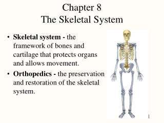
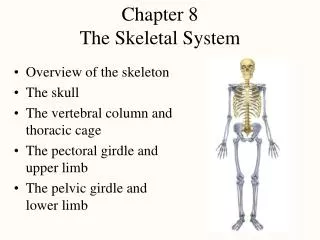
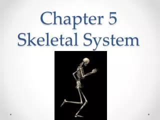
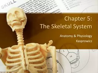
Chapter 8 Skeletal System. Introduction. The skeletal system supports the weight of the body, supports and protects body organs, enables the body to move, acts as storage site for minerals, and produces blood cells. Bones: An Overview. Sizes and Shapes

E N D
Introduction • The skeletal system supports the weight of the body, supports and protects body organs, enables the body to move, acts as storage site for minerals, and produces blood cells.
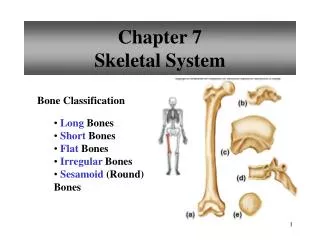
Bones: An Overview • Sizes and Shapes • Bones are classified as long, short, flat, and irregular. • Bone markings function as sites of muscle attachments and passages for nerves and blood vessels. • A long bone has a diaphysis (shaft) and two epiphyses (ends). Articular cartilage is found on the outer surface of the epiphyses. • The diaphysis is composed of compact or hard bone. The epiphysis consists of spongy or soft bone; red marrow is found in the holes of spongy bone.
Bones: An Overview - cont’d • Bone Formation and Growth • Bones ossify in two ways. In the skull, osteoblasts replace thin connective tissue membrane, forming flat bones. Other bones form on hyaline cartilage models as osteoblasts replace cartilage with bone. • Bones grow longitudinally at the epiphyseal disc, to determine height; bones also grow thicker and wider to support the weight of the body. • Bone growth and reshaping occur throughout life and depend on many factors, including diet, exercise, and hormones.
Divisions of the Skeletal System • The names of the 206 bones of the skeleton are listed in Table 8-2.
Divisions of the Skeletal System - cont’d • Axial Skeleton • The axial skeleton includes the bones of the skull (cranium and face), hyoid bone, bones of the middle ear, bones of the vertebral column, and the thoracic cage. • The skull of a newborn contains fontanels, which are membranous areas that allow brain growth. • The skull contains air-filled cavities called sinuses.
Divisions of the Skeletal System - cont’d • Axial Skeleton—cont’d • The vertebral column is formed from 26 vertebrae, one sacrum, and one coccyx. The vertebrae are separated by cartilaginous discs. The vertebral column of the adult has four curvatures: cervical, thoracic, lumbar, and sacral. • The thoracic cage is a bony, cone-shaped cage formed by the sternum, 12 pairs of ribs, and thoracic vertebrae.
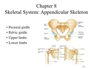
Divisions of the Skeletal System - cont’d • Appendicular Skeleton • The appendicular skeleton includes the bones of the extremities (arms and legs), and the bones of the hip and shoulder girdles. • The shoulder girdle consists of the scapula and the clavicle. • The pelvic girdle is formed by the two coxal bones and is secured to the axial skeleton at the sacrum.
Joints • A joint or articulation is the site where two bones meet.
Joints - cont’d • Types of Joints (based on the degree of movement) • Immovable joints. • Slightly movable joints. • Freely movable joints or synovial joints. Structures within a synovial joint (knee): articular cartilage, the joint capsule, synovial membrane, synovial fluid, bursae, and supporting ligaments. • The types of freely movable joints include hinge, ball and socket, pivot, gliding, saddle, and condyloid.
Joints - cont’d • Joint Movement • Freely movable joints are capable of different types of movement. • Types of movements at freely movable joints include flexion and extension, abduction and adduction, inversion and eversion, supination and pronation, and circumduction.
Introduction • The integumentary system includes the skin, which covers the body, protects the internal organs, and plays an important role in the regulation of body temperature.
Structures: Organs of the Integumentary System • The integumentary system includes the skin, accessory structures, and subcutaneous tissue beneath the skin.
Structures: Organs of the Integumentary System - cont’d • Skin • The skin is called the cutaneous membrane. • The skin has two layers, an outer layer called the epidermis and an inner layer called the dermis. • The epidermis has five layers. The stratum germinativum is the layer in which cell division takes place. The new cells produce keratin (waterproofing) and die as they are pushed toward the surface. The outer layer is the stratum corneum and consists of flattened, dead, keratinized cells.
Structures: Organs of the Integumentary System - cont’d • Skin—cont’d • The dermis lies on the subcutaneous tissue. • Skin color is determined by many factors: some genetic, some physiologic, and some due to disease. Melanin causes skin to darken. Carotene causes skin to appear yellow. The amount of blood in the skin affects skin color (e.g., flushing) as does the appearance of abnormal substances such as bilirubin (jaundice) and a low blood oxygen content (cyanosis).
Structures: Organs of the Integumentary System - cont’d • Accessory Structures of the Skin • Hair is unevenly distributed over the skin. The location of the hair determines its function. Eyebrows and eyelashes protect the eyes from dust and perspiration. • The main parts of a hair are the shaft and root. • Hair color is determined by the amount and type of melanin. • Nails are thin plates of stratified squamous epithelial cells that contain a hard form of keratin. • There are two major exocrine glands in the skin: sebaceous glands and sweat glands.
Structures: Organs of the Integumentary System - cont’d • Accessory Structures of the Skin—cont’d • The sebaceous glands (oil glands) secrete sebum. The sebum lubricates hair and skin. In the fetus, these glands secrete vernix caseosa, a cheeselike substance that coats the skin of a newborn. • The two types of sweat glands (sudoriferous glands) are the apocrine glands and the eccrine glands. The eccrine sweat glands play a crucial role in temperature regulation. • The mammary glands (which secrete milk) and the ceruminous glands (which secrete ear wax) are modified sweat glands.
Structures: Organs of the Integumentary System - cont’d • Subcutaneous Tissue • Subcutaneous tissue anchors the dermis to underlying structures. • Subcutaneous tissue acts as an insulator; it prevents heat loss.
Regulation of Body Temperature • Heat Production • Heat produced by metabolizing cells constitutes the body temperature. • Most of the heat is produced by the muscles and the liver. • Heat Loss • Most of the heat (80%) is lost through the skin. • Heat loss occurs through radiation, conduction, convection, and evaporation.
Regulation of Body Temperature - cont’d • Heat Loss—cont’d • Normal body temperature is set by the body’s thermostat in the hypothalamus. • Heat is lost through sweating and vasodilation. Heat is conserved by vasoconstriction and produced by shivering.
When Skin Is Burned • Physiological Effects • Short-term effects: fluid and electrolyte losses, shock, inability to regulate body temperature, infection • Long-term effects: scarring, loss of function, and cosmetic and emotional problems • Classification of Burns • Classified according to the thickness of the burn (partial, full); also first, second, and third degree. • The rule of nines is a way to evaluate burns.
Introduction • Tissues are groups of cells similar to each other in structure and function. • Membranes are thin sheets of tissue that cover surfaces, line body cavities, and surround organs.
Types of Tissue • Epithelial Tissue Types • Epithelial tissue covers surfaces, lines cavities, and engages in secretion/absorption and protective functions. • Epithelial tissue is classified according to cell shape (squamous, cuboidal, and columnar) and layers (simple and stratified). • The types and functions are summarized in Table 6-1.
Types of Tissue - cont’d • Connective Tissue • The primary function of connective tissue is to bind together the parts of the body. Other functions include support, protection, fat storage, and transport of substances. • Connective tissue has an abundant intercellular matrix that fills spaces between cells. The intercellular matrix may be liquid, gel-like, or hard. The matrix often contains protein fibers that are secreted by the cells. • There are three types of loose connective tissue: areolar, adipose, and reticular.
Types of Tissue - cont’d • Connective Tissue—cont’d • Dense fibrous connective tissue forms tendons, ligaments, capsules, and fascia, and is found in the skin (dermis). • Types of cartilage include: hyaline, elastic, and fibrocartilage. • Bone (osseous tissue) is connective tissue formed by osteocytes. Bone cells have a hard intercellular matrix that includes collagen, calcium salts, and other minerals. • Blood and lymph are types of connective tissue that have a watery intercellular matrix.
Types of Tissue - cont’d • Nervous Tissue • Nervous tissue is found in the peripheral nerves, brain, and spinal cord. • The two types of nervous tissue are neurons, which transmit electrical signals, and neuroglia, which support and take care of the neurons. • Muscle Tissue • Muscle cells contract, thereby causing movement. • The three kinds of muscle are skeletal, smooth, and cardiac.
Tissue Repair • Tissue Repair by Regeneration • Replacement of tissue by cells that undergo mitosis • Tissue Repair by Fibrosis • Formation of scar tissue
Membranes • Epithelial Membranes • The cutaneous membrane is the skin. • Mucous membranes are epithelial membranes that line all body cavities that open to the exterior of the body. • Serous membranes are epithelial membranes that line the ventral body cavities, which are not open to the exterior of the body. • Serous membranes form two layers: a parietal layer that lines the wall of the cavity and a visceral layer that covers the outside of an organ. • The three serous membranes are the pleura, the pericardium, and the peritoneum.
Membranes - cont’d • Connective Tissue Membranes • Synovial membranes are connective tissue membranes. • Other connective tissue membranes are listed in Table 6-3.