Download
1 / 47
550 likes | 873 Vues
The Skeleton. Bones – An introduction. The human skeleton consists of 206 bones. We are actually born with more bones (about 300), but many fuse together as a child grows up. These bones support your body and allow you to move.

E N D
Bones – An introduction • The human skeleton consists of 206 bones. We are actually born with more bones (about 300), but many fuse together as a child grows up. These bones support your body and allow you to move. • The longest bone in our bodies is the femur (thigh bone). The smallest bone is the stirrup bone inside the ear. Each hand has 26 bones in it. Your nose and ears are not made of bone; they are made of cartilage, a flexible substance that is not as hard as bone.
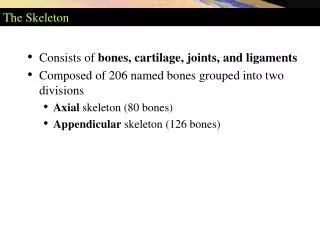
The Axial & Appendicular Skeleton The skeleton is split into these 2 categories: Axial Skeleton – Skull, vertebral column and ribcage Appendicular Skeleton – Limbs, wings and pelvis COMPLETE WORKSHEET 1 C
The Vertebral Column Is there movement? Yes Very limited movement to create a stable structure for the organs found inside the ribcage 2 lumbar vertebrae are moveable, 3 are fused, therefore no movement Fused, therefore no movement
ANSWER THESE QUESTIONS How many bones are there in the human body? What is the longest bone in the body? Where in the body is the smallest bone? Your nose and ears aren’t made of bone but what other substance? The skeleton is defined into two categories, what are they? How many sections make up the vertebral column? 206 femur the ear cartilage axial & appendicular 5
Joints What is a joint? A place where two or more bones meet http://www.youtube.com/watch?v=zWo9- 3GJpr8&feature=PlayList&p=1D1576CEF4196B76&index=5 What are the types of joint? There are 3 types of joint: • Freely Moveable / Synovial • Slightly Moveable / Cartilaginous • Immoveable / Fixed
There are 6 types of freely moveable or synovial joints joints in which we need to know 3 of them: http://www.youtube.com/watch?v=BXoMa2bVC18&feature=related • BALL AND SOCKET JOINTS • HINGE JOINTS • PIVOT JOINT • CONDYLOID- AS IN WRIST • GLIDING- AS IN ANKLE AND HAND • SADDLE- AS IN THUMB
Activity Write down 5 sports actions and beside each write down the main parts of the body that Move, the bones and the type of joint and joint action and movement possible for that action E.g. Snooker. Cueing the white ball. Right arm humerus, ulna, radius, carpal, metacarpal, phalanges. Elbow, synovial, hinge lots of movement
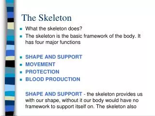
Function of the Skeleton – worksheet 1e • Your skeleton has many different functions to enable you to live and survive. These include: • Protection • Movement • Shape • Muscle/organ attachment • Blood cell production • bone growth • http://www.youtube.com/watch?v=zZxLfJRd4cs
Protection – Many of the internal organs in your body are protected by bone. The flat type bones of your body function in this way (sternum, pelvis, cranium etc.) • Movement – Your body is able to move because of the co-operation between its muscles and bones. The muscles are attached to the bones of the skeleton, creating a lever and joints system that allows the body to move. • Shape – Your skeleton provides the framework to give your body its shape. Without it, you would look like a blob of jelly.
Muscle/organ – Your body provides a support system to attach muscles, organs, arteries, veins etc. • Blood cell production – Bone also produces blood cells. This occurs in the marrow of the bone that is found in the epiphysis (end) and diaphysis (shaft) of all long bones. • Bone growth – As we discovered earlier bone has the ability to be broken down and reformed. This is called ossification.
http://www.bbc.co.uk/science/humanbody/body/index_interactivebody.shtmlhttp://www.bbc.co.uk/science/humanbody/body/index_interactivebody.shtml
Bone Growth Bones start off as cartilage as a fetus. As the fetus develops, mineral are laid down and the cartilage becomes harder and less flexible. This process is called ‘ossification’ and can continue until you are 30 (although it usually stops between 18-21) Bone is continually being broken down and replaced; this process is done by 2 different cells: Osteoclasts break down old bone and clean the bone environment. Osteoblasts are bone forming cells that help to develop new bone throughout life. These will replace about 10% of bone every year; this means that no matter how old we are our skeleton is no older than 10 years old.
Structure of a Long Bone Epiphysis – Ends of the bone. Diaphysis – Long shaft of the bone. Articular cartilage – thin layer of blueish cartilage covering each end of the bone. Periosteum – thin outer layer of the bone. It contains nerves and blood vessels that feed the bone. Compact bone – This is hard and resistant to bending. Spongy bone – this lies in layers within the compact bone. It has a honeycomb appearance and gives bones their elastic strength. Medullary cavity – the hollow space down the middle of the compact bone and contains bone marrow. Bone marrow produces blood cells and store fat.
HOW DO WE CLASSIFY BONES ? Bones are classified according to their function. • (Protection) FLAT BONES • (Protection) IRREGULAR BONES • (Levers) LONG BONES • (Small movements)SHORT BONES
Effects of Exercise The skeletal system changes due to exercise. However, the changes depend on the type of exercise that individuals may participate in. The changes can be short or long term: Short term: When you participate in exercise your body moves more rapidly which means that joints need to work more. This extra demand on the joints causes a release of synovial fluid around the joint site that helps movement occur more easily. Long term: Hyaline cartilage increases in its thickness around the joint site as exercise continues. This can help with preventing the surface of the bones from wearing away too soon. Regular participation in weight bearing exercises alsohelps to increase bone density, resulting in the bones becoming stronger.
Describing the body’s parts and position Anterior – Front of body Posterior – Back of body Midline Superior Superior – closer to head Inferior – further away from head Inferior Medial – closer to midline of body Lateral – further away from midline of the body Lateral Proximal Medial Proximal – nearer point of attachment of main structure of body Distal – further away from point of attachment to the main structure of the body Distal Anterior Posterior