Download
1 / 6
60 likes | 70 Vues
Rotavirus (RV) is the most common cause of severe<br>dehydrating diarrhoea in healthy infants and young children.<br>The aims of this study were to investigate a RV outbreak in the<br>pediatric hematology and oncology ward and to examine possible<br>associations between immune status and RV infection.

E N D
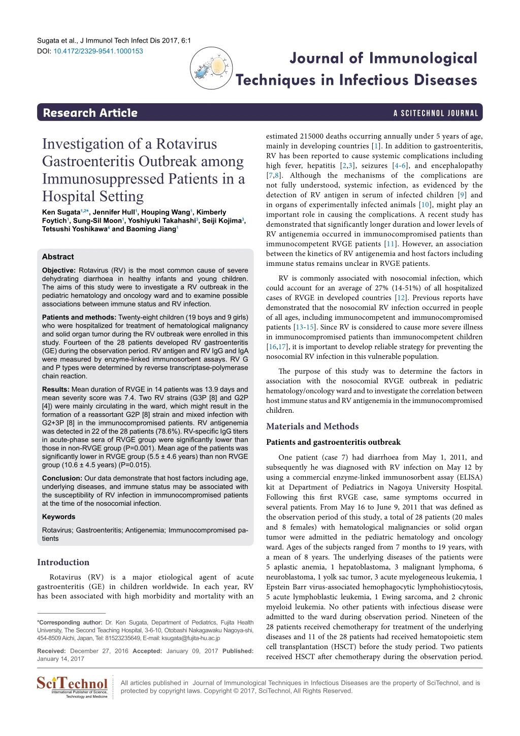
Sugata et al., J Immunol Tech Infect Dis 2017, 6:1 DOI: 10.4172/2329-9541.1000153 Journal of Immunological Techniques in Infectious Diseases Research Article a SciTechnol journal estimated 215000 deaths occurring annually under 5 years of age, mainly in developing countries [1]. In addition to gastroenteritis, RV has been reported to cause systemic complications including high fever, hepatitis [2,3], seizures [4-6], and encephalopathy [7,8]. Although the mechanisms of the complications are not fully understood, systemic infection, as evidenced by the detection of RV antigen in serum of infected children [9] and in organs of experimentally infected animals [10], might play an important role in causing the complications. A recent study has demonstrated that significantly longer duration and lower levels of RV antigenemia occurred in immunocompromised patients than immunocompetent RVGE patients [11]. However, an association between the kinetics of RV antigenemia and host factors including immune status remains unclear in RVGE patients. Investigation of a Rotavirus Gastroenteritis Outbreak among Immunosuppressed Patients in a Hospital Setting Ken Sugata1,2*, Jennifer Hull1, Houping Wang1, Kimberly Foytich1, Sung-Sil Moon1, Yoshiyuki Takahashi3, Seiji Kojima3, Tetsushi Yoshikawa4 and Baoming Jiang1 Abstract Objective: Rotavirus (RV) is the most common cause of severe dehydrating diarrhoea in healthy infants and young children. The aims of this study were to investigate a RV outbreak in the pediatric hematology and oncology ward and to examine possible associations between immune status and RV infection. RV is commonly associated with nosocomial infection, which could account for an average of 27% (14-51%) of all hospitalized cases of RVGE in developed countries [12]. Previous reports have demonstrated that the nosocomial RV infection occurred in people of all ages, including immunocompetent and immunocompromised patients [13-15]. Since RV is considered to cause more severe illness in immunocompromised patients than immunocompetent children [16,17], it is important to develop reliable strategy for preventing the nosocomial RV infection in this vulnerable population. Patients and methods: Twenty-eight children (19 boys and 9 girls) who were hospitalized for treatment of hematological malignancy and solid organ tumor during the RV outbreak were enrolled in this study. Fourteen of the 28 patients developed RV gastroenteritis (GE) during the observation period. RV antigen and RV IgG and IgA were measured by enzyme-linked immunosorbent assays. RV G and P types were determined by reverse transcriptase-polymerase chain reaction. The purpose of this study was to determine the factors in association with the nosocomial RVGE outbreak in pediatric hematology/oncology ward and to investigate the correlation between host immune status and RV antigenemia in the immunocompromised children. Materials and Methods Patients and gastroenteritis outbreak Results: Mean duration of RVGE in 14 patients was 13.9 days and mean severity score was 7.4. Two RV strains (G3P [8] and G2P [4]) were mainly circulating in the ward, which might result in the formation of a reassortant G2P [8] strain and mixed infection with G2+3P [8] in the immunocompromised patients. RV antigenemia was detected in 22 of the 28 patients (78.6%). RV-specific IgG titers in acute-phase sera of RVGE group were significantly lower than those in non-RVGE group (P=0.001). Mean age of the patients was significantly lower in RVGE group (5.5 ± 4.6 years) than non RVGE group (10.6 ± 4.5 years) (P=0.015). One patient (case 7) had diarrhoea from May 1, 2011, and subsequently he was diagnosed with RV infection on May 12 by using a commercial enzyme-linked immunosorbent assay (ELISA) kit at Department of Pediatrics in Nagoya University Hospital. Following this first RVGE case, same symptoms occurred in several patients. From May 16 to June 9, 2011 that was defined as the observation period of this study, a total of 28 patients (20 males and 8 females) with hematological malignancies or solid organ tumor were admitted in the pediatric hematology and oncology ward. Ages of the subjects ranged from 7 months to 19 years, with a mean of 8 years. The underlying diseases of the patients were 5 aplastic anemia, 1 hepatoblastoma, 3 malignant lymphoma, 6 neuroblastoma, 1 yolk sac tumor, 3 acute myelogeneous leukemia, 1 Epstein Barr virus-associated hemophagocytic lymphohistiocytosis, 5 acute lymphoblastic leukemia, 1 Ewing sarcoma, and 2 chronic myeloid leukemia. No other patients with infectious disease were admitted to the ward during observation period. Nineteen of the 28 patients received chemotherapy for treatment of the underlying diseases and 11 of the 28 patients had received hematopoietic stem cell transplantation (HSCT) before the study period. Two patients received HSCT after chemotherapy during the observation period. Conclusion: Our data demonstrate that host factors including age, underlying diseases, and immune status may be associated with the susceptibility of RV infection in immunocompromised patients at the time of the nosocomial infection. Keywords Rotavirus; Gastroenteritis; Antigenemia; Immunocompromised pa- tients Introduction Rotavirus (RV) is a major etiological agent of acute gastroenteritis (GE) in children worldwide. In each year, RV has been associated with high morbidity and mortality with an *Corresponding author: Dr. Ken Sugata, Department of Pediatrics, Fujita Health University, The Second Teaching Hospital, 3-6-10, Otobashi Nakagawaku Nagoya-shi, 454-8509 Aichi, Japan, Tel: 81523235649, E-mail: ksugata@fujita-hu.ac.jp Received:December 27, 2016Accepted:January 09, 2017Published: January 14, 2017 All articles published in Journal of Immunological Techniques in Infectious Diseases are the property of SciTechnol, and is protected by copyright laws. Copyright © 2017, SciTechnol, All Rights Reserved. International Publisher of Science, Technology and Medicine
Citation: Sugata K, Hull J, Wang H, Foytich K, Moon SS, et al. (2017) Investigation of a Rotavirus Gastroenteritis Outbreak among Immunosuppressed Patients in a Hospital Setting. J Immunol Tech Infect Dis 6:1. doi: 10.4172/2329-9541.1000153 Three of the 11 transplant recipients had diarrhoea during the observation period, which was considered to be acute graft-versus- host disease. In addition, 11 immunosuppressed patients without HSCT also developed diarrhoea during the observation period. Stool samples collected from patients with gastroenteritis were tested for adenovirus by using a commercial ELISA kit (Adenoclone, Meridian Bioscience, Inc., Cincinnati, OH). Adenovirus antigen was detected in only one patient (case 28) and no bacteria were isolated from the samples. wells plates were coated overnight with rabbit hyper immune serum to RV strain RRV at 4°C. After washing and blocking with MPBS for 1 hour at 37°C, plates were incubated with supernatants of RRV- infected MA104 cells (1:10 diluted in diluent buffer) for 1 hour at 37°C. After washing, serum samples that were serially diluted from 1:10 to 1:5120 in diluent buffer were added, followed by a secondary goat anti-human IgG-biotin (1:3000) or human IgA-biotin (1:2000) (KPL Inc., Gaithersburg, MD) in MPBS for 1 hour at 37°C. After washing, extra Vidin (1:3000) (Sigma-Aldrich, St. Louis, MO) was added for 1 hour at 37°C. After adding the substrate, OD was read at 450 nm. IgG and IgA titers in serum were calculated as the reciprocal of the highest dilution that gave a mean OD greater than the cut-off value (3 standard deviations above the mean OD of the negative- control serum wells). G and P typing and sequence analysis: G genotypes of RV in stool were determined by using reverse transcription polymerase chain reaction (RT-PCR) with previously published specific primers 9con1L and VP7-RDg [21]. The second amplification was performed from the first PCR product (1025 bp) using the 9con1L primer and a cocktail of G type- specific primers (9T-1, 9T-2, 9T-3P, 9T-4 and 9T- B) for VP7 G1 (158 bp), G2 (224 bp), G3 (466 bp), G4 (403 bp) and G9 (110 bp), respectively. Clinical manifestations were examined retrospectively from medical records. The severity of GE was evaluated by using a 20-point Vesikari scoring scale [18]. Laboratory findings including white blood cell (WBC), neutrophil and lymphocyte counts were examined on May 9 in all subjects for evaluation of the host immune status. Informed consent was obtained from patient’s parents before enrollment of this study. This study was approved by the Ethics Committee of Fujita Health University School of Medicine. This study did not require review by the Center for Disease Control (CDC) Institutional Review Board because the CDC tested pre-existing, anonymous specimens. Sample collection Serum samples were collected weekly for one month of the observation period from 25 patients and collected for two weeks from 3 patients who were discharged earlier from the hospital. Stool specimens were collected weekly from 14 patients with gastrointestinal symptoms. The rectal swabs were kept in 10% phosphate buffered saline (PBS). A total of 106 serum and 44 rectal swab samples were collected from the patients and 5 serum samples were also collected from age-matched healthy children as control. All samples were stored at -70 ºC before shipping for analysis of the Viral Gastroenteritis. Laboratory of the US CDC ELISA for the detection of rotavirus antigenemia: RV antigen in stool and serum was measured using an in-house ELISA specific for RV VP6 [19]. To prevent potential cross-contamination of samples, stool and serum were prepared in different rooms. Briefly, 96 well plates (Nalge Nunc International, Rochester, NY) coated with a monoclonal antibody against the VP6 antigen of RV (YO-156) were blocked with 1% bovine serum albumin in PBS containing Tween 20 (PBST), washed with PBST, then incubated with 50 μl of stool extract (10% in PBS) or diluted serum (1:8 in PBS) at 4°C overnight. After washing, 50 µl of rabbit anti-human RV hyper immune serum diluted 1:5,000 with PBST containing 2.5% skim milk (MPBS) were added at 37°C for 1.5 hours. After washing, a 1:2,000 dilution of peroxidase conjugated donkey anti-rabbit IgG (Jackson Immuno Research Laboratory Inc., West Grove, PA) were added at 37°C for 1.5 hours. After adding the substrate, the optical density (OD) was read at 450 nm with an EIA reader (MRX Revelation, Dynex Technologies, Chantilly, VA). As the mean OD of the control samples was 0.142±0.017, we defined 0.194 (mean+3 SD) as the baseline value in this study. ELISA for the detection of rotavirus antibody: To evaluate role of the immune status in RV infection, RV IgA and IgG titers were examined in acute (1st week of the observation period) and convalescent sera (4th week of the observation period) collected from all hospitalized patients except for cases 26, 27 and 28. Serum collected in 2nd week was used as convalescent phase sample in the three patients because they were discharge before 4th week. Antibody titers were measured by ELISAs for RV IgG and IgA [20]. Briefly, 96 RV VP4 P typing was performed in the same manner as VP7 gene assay with previously published specific primers Con2 and Con3 [22]. The second amplification was performed from the first PCR product (877 bp) using the Con3 primer and a cocktail of P type-specific primers (2T-1, 3T-1 and 1T-1) for P4 (484 bp), P6 (268 bp) and P8 (346 bp), respectively. The PCR products of rotavirus VP7 and VP4 genes were purified by mini columns (QIAquick, Qiagen, Valencia, CA) and sequences were determined by using the ABI-PRISM Big Dye terminator Cycle Sequencing kit and an ABI Prism 310 Genetic analyzer (Applied Biosystems Inc. Foster City, CA). Statistical analysis Statistical analyses among clinical variables, RV antigen levels and immunoglobulin levels in serum were performed using SPSS version 20. The one way ANOVA analysis of variance by ranks procedure was employed to test mean severity scores and WBC, neutrophil, lymphocyte cell counts and duration of the admission among the patients, followed by pair-wise examinations using the Mann-Whitney U test for unpaired data. Levels of RV antigenemia and titers of RV IgA and IgG in serum of patients with or without gastroenteritis were compared using either a paired Wilcoxon signed ranks test or Mann-Whitney U test. Gender, underlying diseases, chemotherapy of patients with or without gastroenteritis during study period and HCST prior to this study were compared using Fisher’s exact probability test. In all comparisons, a p-value<0.05 (two-tailed) was considered statistically significant. Correlations among the duration of GE, RV antigen in serum samples, RV antigen in stool, total Vesikari severity score, and individual symptom scores were examined using Spearman rank correlation coefficient. Results RV antigen in serum and stool Of the 28 patients admitted for chemotherapy in the hospital, one half had developed diarrhoea during their stay. These 14 GE patients had a mean duration of 13.9 days and a mean Vesikari severity score of 7.4. RV antigen was detected in 29 (65.9%) of the 44 stool samples • Page 2 of 6 • Volume 6 • Issue 1 • 1000153
Citation: Sugata K, Hull J, Wang H, Foytich K, Moon SS, et al. (2017) Investigation of a Rotavirus Gastroenteritis Outbreak among Immunosuppressed Patients in a Hospital Setting. J Immunol Tech Infect Dis 6:1. doi: 10.4172/2329-9541.1000153 collected from the 14 patients with GE and in 69 (65.1%) of the 106 serum samples from 22 of the 28 patients (except cases 4, 5, 9, 19, 22, and 27) (Figure 1). Of the 22 patients with RV antigenemia, 11 had gastroenteritis (cases 3, 7, 8, 10, 11, 12, 15, 23, 24, 26, and 28) and the other 11 did not (cases 1, 2, 6, 13, 14, 16, 17, 18, 20, 21, and 25). RV antigenemia lasted for entire observational period (4 weeks) in 12 of the 22 antigenemia-positive patients (cases 1, 2, 6, 7, 10, 11, 14, 15, 20, 21, 23, and 24). RV antigenemia was not observed in the 3 patients with positive RV antigen in stool (cases 19, 22, and 27). Serological analysis RV IgG titer in acute phase was significantly lower in RVGE group 1 2 3 4 1 2 3 4 1 2 3 4 (weeks) case case case 21 1 11 2 12 22 3 13 23 4 14 24 5 15 25 6 16 26 7 17 27 8 18 28 9 19 10 20 Figure 1: Associations between rotavirus antigens in stool (black boxes) and rotavirus antigenemia (shaded bars). White boxes indicate rotavirus antigen negative in both stool and serum samples. Stool samples were collected only if the patient had GE symptoms. Case 26 died of the underlying disease and case 27 and 28 patients were discharged during the study period. • Page 3 of 6 • Volume 6 • Issue 1 • 1000153
Citation: Sugata K, Hull J, Wang H, Foytich K, Moon SS, et al. (2017) Investigation of a Rotavirus Gastroenteritis Outbreak among Immunosuppressed Patients in a Hospital Setting. J Immunol Tech Infect Dis 6:1. doi: 10.4172/2329-9541.1000153 A. RV GE group (n=14) non RV GE group (n=14) 10000 P= 0.001 Serum IgG titer 1000 100 10 1 1st week 1st week non RVGE group (n=14) B. RVGE group (n=14) C. P= 0.022 P= 0.310 Serum IgG titer 4th week 1st week 4th week 1st week Figure 2: Antibody profiles in sera of immunosuppressed patients. A. Comparison of rotavirus-specific IgG titers in acute sera from RVGE patients and non RVGE patients (samples collected in the 1st week of the outbreak). B. B and C. Kinetics of rotavirus-specific IgG titers from acute phase (1st week of the outbreak) to convalescent phase (4th week) of RVGE and non RVGE patients. (median, 345, range: 40–2560) than non GE group (median, 3800, range: 80–20480) (P=0.001) (Figure 2A). However, no significant difference was observed in acute phase IgA antibody titers between RVGE patients (median, 117, range: 10–320) and non GE patients (median 87, range: 40–160) (P=0.910). Antibody titers were compared in paired sera obtained from patients with and without RVGE. A significant increase in RV IgG titers was demonstrated in convalescent phase samples (median, 2565 fold, range: 80–20480) in comparison to acute phase samples (median, 345 fold, range: 40–2560) from RVGE patients (P=0.022) in RVGE group (Figure 2B). In contrast to RVGE group, no significant difference was observed between acute phase RV IgG antibody titers (median 3800 fold, range: 80–20480) and convalescent phase one(median 4868 fold, range: 320–20480) (P=0.310) in non RVGE group (Figure 2C). No significant difference was observed in RV IgA antibody titers between acute-phase samples and convalescent-phase samples in either RVGE or non-RV patients (date not shown). Factors in association with RVGE Patients’ characteristics and clinical features were compared between the RVGE patients and non-RVGE patients to elucidate factors in association with RVGE in these subjects (Table 1). Mean age of the patients was significantly lower in RVGE group (5.5±4.6 years) than non RVGE group (10.6 ± 4.5 years) (P=0.015). Gender was not associated with RVGE (P=0.103). Neither HSCT (P=1.0) nor chemotherapy (P=1.0) was associated with RVGE. No significant difference in WBC (P=0.401), neutrophil counts (P=0.511), and lymphocyte counts (P=0.352) was demonstrated between the two groups. There was no significant difference in the levels of mean (P=0.769) and maximal (P=0.872) serum RV antigen levels between 11 RVGE patients and 11 non GE patients. Molecular epidemiological analysis in RVGE outbreak RV antigen was detected in 29 of 44 fecal swab samples by ELISA. G and P types were determined in 12 of the 29 samples by using RT- PCR. Two strains (G3P [8] (4/17, 23.5%) and G2P [4] (4/17, 23.5%) were dominant in the analyzed samples, followed by G2P [8] (2/17, 11.8%) and mixed G2/G3P [8] (2/17, 11.8%). Discussion Although it has been demonstrated that RV can cause severe clinical manifestations in immunocompromised transplant recipients [23-25], few studies have been conducted to examine the full • Page 4 of 6 • Volume 6 • Issue 1 • 1000153
Citation: Sugata K, Hull J, Wang H, Foytich K, Moon SS, et al. (2017) Investigation of a Rotavirus Gastroenteritis Outbreak among Immunosuppressed Patients in a Hospital Setting. J Immunol Tech Infect Dis 6:1. doi: 10.4172/2329-9541.1000153 the emergence of new reassortant strains, and the outbreak of this nosocomial infection. On the other hand, we demonstrated an equally high prevalence of antigenemia in immunocompromised patients with or without RVGE, suggesting that antigenemia may not contribute the expression of GE. RV strain G1P8 was the most predominant and followed by G2P [4] and G3P [8] between 1994 and 2003 in the world [31]. It has been demonstrated that introduction of RV vaccines affected the prevalence of endemic strains in some Asian countries [32,33]. Additionally, G9P [8], G2P [4], G3P [8], G1P [4], and G9P [6] were also detected during the period [34]. Then, G3P [8] strain emerged (65.0%) during 2010-2011 season, and G2P [4] strain was rarely detected (2.4%) at that time period [35]. This outbreak occurred just before introduction of RV vaccine in Japan. Two types of RV strains G3P [8] and G2P [4] were mainly circulating in this outbreak, which resulted in nosocomial RVGE in almost half of the immunocompromised patients in a pediatric hematology/oncology ward (Table 2). It is likely that G3P [8] strain invaded into the ward before G2P [4] invasion. As G2P [8] has been reported to be uncommon among healthy children in several developed countries including Japan [35-38], we thought that strain G2P [8] identified in this outbreak would be a reassortant between G3P [8] and G2P [4] strains. spectrum of RV infection in these patients. Stelzmueller et al. [23] demonstrated that RV infection was observed in 1.5% of solid organ transplant recipients, and the highest frequency of RV infections was observed in pediatric liver transplant recipients (52%) based on conventional RV antigen detection analysis of stool samples. RV infection has also been identified in 10-12% of pediatric bone marrow transplant recipients [26,27]. In this outbreak, GE was observed in 14 of the 28 (50.0%) patients. All diarrhoeal stool samples collected from 14 GE patients were positive for RV antigen. In addition, eleven asymptomatic patients with RV antigenemia were identified in this cohort. To our knowledge, this is the first study demonstrating a high prevalence of RV antigenemia in a nosocomial RVGE outbreak among immunosuppressed patients. Persistent RV antigenemia observed in this outbreak corresponded with our previous data in transplant patients [12]. Future studies are needed to elucidate clinical implications of persistent low level RV antigenemia in immunocompromised patients. It has been demonstrated that RV infection was more severe in transplant recipients than immunocompetent patients [28]. In the present study, we observed a long duration of GE (13.9 days) but a relatively low mean Vesikari severity score of 7.4. This low severity of GE in this cohort might be due to older age (5.5 years) of the patients. It is highly likely that these patients had RV infection before, which is known to protect against subsequent infection and severity of RVGE [29,30]. Of note, the patients who did not develop RVGE had a significantly higher mean age than those in RVGE group in this outbreak. Our present study has several limitations. First, we only enrolled a relatively small number of transplant patients in one hospital for four weeks. Second, we did not collect stool samples from non GE patients, hospital staff or patients’ family members. In addition, except for RV and adenovirus we did not look for other viral pathogens that might cause this GE outbreak as well. Although it was too small to carry out multivariate regression analysis in this study, younger patient and frequent movement to other wards were apparent key risk factors for nosocomial RV infection in pediatric hematology/oncology ward. High prevalence of RV infection in transplant patients and patterns of RV transmission in hospital wards are useful information which should encourage scientists and clinicians to develop measures and strategies to manage and protect this vulnerable population in health care settings. From clinical perspectives, it is very important to elucidate risk factors for RVGE in order to better prevent nosocomial infection among immunocompromised patients in the ward. Our present study found several possible risk factors for RVGE in this setting. First, lower titer of RV IgG antibody and younger age appeared to be associated with RVGE in patients. Second, tracking of the patients’ movements and characterization of RV strains from patients in the same or nearby wards helped us determine that the transfer of patients between the wards might have contributed to the spread of common rotavirus strains, Acknowledgments Potential conflicts of interest. All authors report no conflicts of interest relevant to this article. Rotavirus gastroenteritis Yes (n=14) 5.5 ± 4.6 Categories P Financial Support: none reported. No (n=14) 10.6 ± 4.5 Age (year) Gender Male Female HSCT Done ND Chemotherapy Receiving Not receiving 0.015* Case numbers 7 Date of onset May 1st May 16th May 23rd May 30th June 6th 12 2 7 7 0.103 G3P[8] G2P[NT] G2/3P[8] G2P[8] 10 May 8th 5 6 8 May 9th G3P[8] G3P[8] G3P[8] 1.000 9 8 12 May 9th G2P[8] 11 May 12th G2P[4] 8 9 1.000 22 May 12th G2/3P[8] 6 5 23 May 12th G2P[4] WBC (×109/L) 2.4 ± 1.7 4.0 ± 4.5 0.401 0.511 24 May 12th G2P[NT] G2P[NT] 26 May 12th Neutrophil 1.3 ± 1.3 2.5 ± 3.7 28 May 12th G2P[4] Lymphocyte RV antigenemia Mean OD Maximal OD 0.83 ± 5.6 (n=11) 0.246 ± 0.067 0.266 ± 0.070 1.3 ± 1.1 (n=11) 0.250 ± 0.096 0.274 ± 0.103 0.352 0.769 19 May 13th G2P[NT] 3 May 14th G2P[NT] G2P[4] 27 May 14th G[NT]P[NT] 0.872 15 May 17th G[NT]P[NT] Movement (time) 2.9 ± 1.4 1.9 ± 1.0 0.039* Table 2: Onset of diarrhoea and RV strains. RV strains Table 1: Characteristics and clinical features of the patients. • Page 5 of 6 • Volume 6 • Issue 1 • 1000153
Citation: Sugata K, Hull J, Wang H, Foytich K, Moon SS, et al. (2017) Investigation of a Rotavirus Gastroenteritis Outbreak among Immunosuppressed Patients in a Hospital Setting. J Immunol Tech Infect Dis 6:1. doi: 10.4172/2329-9541.1000153 Rotavirus enteritis in solid organ transplant recipients: an underestimated problem? Transpl Infect Dis 9: 281-285. References 1. Tate J E, Burton A H, Boschi-Pinto C, Parashar U D (2016) WHO-Sureveillance Network. Global, Regional, and National Estimates of Rotavirus Mortality in Children <5 Years of Age, 2000-2013. Clin Infect Dis 62: 96-105. 24. Berger N, Wirmsberger R, Kafka R, Margreiter C, Ebenbichler C, et al. (2006) Infectious complications following 72 consecutive enteric-drained pancreas transplants. Transpl Int 19: 549-557. 2. St Geme JW 3rd, Hyman D (1988) Hepatic injury during rotavirus infections. J Pediatr 113: 952-3. 25. Stelzmueller I, Dunst KM, Hengster P, Wykypiel H, Steurer W et al. (2005) A cluster of rotavirus enteritis in adult transplant recipients. Transpl Int 18: 470-474. 3. Teitelbaum JE, Daghistani R (2007) Rotavirus Causes Hepatic Transaminase Elevation. Dig Dis Sci 52: 3396-3398. 26. Cox GJ, Matsui SM, Lo RS, Hinds M, Bowden RA et al. (1994) Etiology and outcome of diarrhoea after marrow transplantation: a prospective study. Gastroenterology 107: 1398–1407. 4. Contino MF, Lebby T, Arcinue EL (1994) Rotaviral gastrointestinal infection causing afebrile seizures in infancy and childhood. Am J Emerg Med 12: 94-95. 27. Troussard X, Bauduer F, Gallet E, Freymuth F, Boutard P et al. (1993) Virus recovery from stools of patients undergoing bone marrow transplantation. Bone Marrow Transplant 12: 573–576. 5. Kawano G, Oshige K, Syutou S, Koteda Y, Yokoyama T, et al. (2007). Benign infantile convulsions associated with mild gastroenteritis: A retrospective study of 39 cases including virological tests and efficacy of anticonvulsants. Brain Dev 29: 617-622. 28. Saulsbury FT, Winkelstein JA, Yolken RH (1980) Chronic rotavirus infection in immunodeficiency. J Pediatr 97: 61-65. 6. Chung B, Wong V (2007). Relationship between five common viruses and febrile seizure in children. Arch Dis Child. 92: 589-593. 29. Velázquez FR, Matson DO, Calva JJ, Guerrero L, Morrow AL, et al. (1996) Rotavirus infection in infants as protection against subsequent infections. N Engl J Med 335: 1022-1028. 7. Kirton A, Busche K, Ross C, Wirrell E (2005). Acute necrotizing encephalopathy in caucasian children: two cases and review of the literature. J Child Neurol 20: 527-532. 30. Fischer TK, Valentiner-Branth P, Steinsland H, Perch M, Santos G, et al. (2002) Protective immunity after natural rotavirus infection: a community cohort study of newborn children in Guinea-Bissau, West Africa. J Infect Dis 186: 593-597. 8. Hongou K, Konishi T, Yagi S, Araki K, Miyawaki T (1998) Rotavirus encephalitis mimicking afebrile benign convulsions in infants. Pediatr Neurol 18: 354-357. 9. Sugata K, Taniguchi K,Yui A, Miyake F, Suga S, et al. (2008) Analysis of Rotavirus Antigenemia and Extra intestinal Manifestations in Children with Rotavirus Gastroenteritis. Pediatrics 122: 392-397. 31. Gentsch JR, Laird AR, Bielfelt B, Griffin DD, Banyai K, et al. (2005) Serotype diversity and reassortment between human and animal rotavirus strains: Implications for rotavirus vaccine programs. J Infect Dis 192: 146-159. 10. Crawford SE, Patel DG, Cheng E, Berkova Z, Hyser JM, et al. (2006) Rotavirus viremia and extra intestinal viral infection in the neonatal rat model. J Virol 80: 4820-4832. 32. Yen C, Tate JE, Patel MM, Cortese MM, Lopman B et al. (2011) Rotavirus vaccines: Update on global impact and future priorities. Hum Vaccin 7: 1282- 1290. 11. Sugata K, Taniguchi K, Yui A, Nakai H, Asano Y, et al. (2012) Analysis of rotavirus antigenemia in hematopoietic stem cell transplant recipients. Transpl Infect Dis 14: 49-56. 33. Phua KB, Lim FS, Lau YL, Nelson EA, Huang LM, et al. (2009) Safety and efficacy of human rotavirus vaccine during the first 2 years of life in Asian infants: randomized, double-blind, controlled study. Vaccine 27: 5936-5941. 12. Fischer TK, Bresee JS, Glass RI (2004) Rotavirus vaccines and the prevention of hospital-acquired diarrhea in children. Vaccine 22: 49-54. 34. Dey SK, Thongprachum A, Ota Y, Phan TG, Nishimura S, et al. (2009) Molecular and epidemiological trend of rotavirus infection among infants and children in Japan. Infect Genet Evol 9: 955-961. 13. Kim CR, Oh JW, Yum MK, Lee JH, Kang JO (2009) Rotavirus infection in neonates at a university hospital in Korea. Infect Control Hosp Epidemiol 30: 893-895. 35. Thongprachum A, Chan-it W, Khamrin P, Okitsu S, Nishimura S, A et al (2013) Reemergence of new variant G3 rotavirus in Japanese pediatric patients, 2009-2011. Infect Genet Evol 13: 168-174. 14. Rodriguez-Baez N, O’Brien R, Qiu SQ, Bass DM (2002) Astrovirus, adenovirus, and rotavirus in hospitalized children: Prevalence and association with gastroenteritis. J Pediatr Gastroenterol Nutr 35: 64-68. 36. Tatte VS, Chitambar SD (2012) Evidence of discordant genetic linkage in the VP4, VP6, VP7 and NSP4 encoding genes of rotavirus strains from adolescent and adult patients with acute gastroenteritis. Infect Genet Evol 12: 1630-1634. 15. Bruijning-Verhagen P, Quach C, Bonten M (2011) Nosocomial rotavirus infections: a meta-analysis. Pediatrics 129: 1011-1019. 37. Bourdett-Stanziola L, Ortega-Barria E, Espinoza F, Bucardo F, Jimenez C, et al. (2010) Rotavirus genotypes in Costa Rica, Nicaragua, Honduras and the Dominican Republic. Intervirology 53: 390-393. 16. Rayani A, Bode U, Habas E et al. (2007) Rotavirus infections in paediatric oncology patients: a matched-pairs analysis. Scand J Gastroenterol 42: 81-87. 17. Mori I, Matsumoto K, Sugimoto K, Kimura M, Daimon N et al. (2002) Prolonged shedding of rotavirus in a geriatric inpatient. J Med Virol 67: 613-615. 38. Westerman LE, McClure HM, Jiang B, Almond JW, Glass RI (2005) Serum IgG mediates mucosal immunity against rotavirus infection. Proc Natl Acad Sci USA 102: 7268-7273. 18. Ruuska T, Vesikari T. (1990) Rotavirus disease in Finnish children: use of numerical scores for clinical severity of diarrhoeal episodes. Scand J Infect Dis 22: 259-267. 19. Taniguchi K, Urasawa T, Urasawa S, Yasuhara T (1984) Production of subgroup-specific monoclonal antibodies against human rotaviruses and their application to an enzyme-linked immunosorbent assay for subgroup determination. J Med Virol 14: 115–125. 20. Moon S. S., Wang Y, Shane A. L, Nguyen T, Ray P, P et al. (2010) Inhibitory effect of breast milk on infectivity of live oral rotavirus vaccines. Pediatr Infect Dis J 29: 919-923. Author Affiliations Top 21. Das BK, Gentsch JR, Cicirello HG, Woods PA, Gupta A et al. (1994) Characterization of rotavirus strains from newborns in New-Delhi, India. J Clin Microbiol 32: 1820–1822. 1Division of Viral Diseases, Centers for Disease Control and Prevention, CDC, Atlanta, GA, USA 2Department of Pediatrics, Fujita Health University, The Second Teaching Hospital, 3-6-10,Otobashi Nakagawaku Nagoya-shi, 454-8509 Aichi, Japan 22. Gentsch JR, Glass RI, Woods P, Gouvea V, Gorziqila M et al. (1992) Identification of group A rotavirus gene 4 types by polymerase chain reaction. J Clin Microbiol 30: 1365-1373. 3Nagoya University, Nagoya, Aichi, Japan 4Fujita Health University, Aichi, Japan 23. Stelzmueller I, Wiesmayr S, Swenson BR, Biebl M, Goegele H et al. (2007) • Page 6 of 6 • Volume 6 • Issue 1 • 1000153