LAPAROSCOPIC SURGERY
LAPAROSCOPIC SURGERY. Jane P Bradley Hendricks RGN, BSE(hons), MCS, Independent Nurse Prescriber Surgical Care Practitioner, Laparoscopic Surgery. Colchester General Hospital. SHORT HISTORY. 1982 Semm performed first Laparoscopic Appendicectomy.


LAPAROSCOPIC SURGERY
E N D
Presentation Transcript
LAPAROSCOPIC SURGERY Jane P Bradley Hendricks RGN, BSE(hons), MCS, Independent Nurse Prescriber Surgical Care Practitioner, Laparoscopic Surgery. Colchester General Hospital
SHORT HISTORY • 1982 Semm performed first Laparoscopic Appendicectomy. • 1987 Mouret performed first Laparoscopic Cholecystectomy. • 1992 First UK Laparoscopic Training centre established.
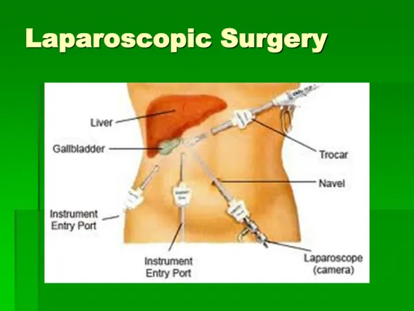
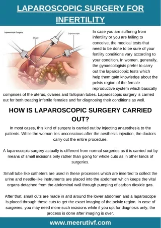
LAPAROSCOPIC SURGERY “KEYHOLE SURGERY” MINIMALLY INVASIVE SURGERY MINIMAL ACCESS SURGERY
Diagnosis Crohn’s Disease Diverticulitis Rectal Prolapse Benign renal disease Gastric Obstruction Some Splenic disorders Operation Bowel resection Bowel resection Repair of Prolapse Nephrectomy Bypass Spleenectomy What operations can we do laparoscopically?
Diagnosis Gallstone Appendicitis Hernia Adhesions Perforated ulcer Hiatus Hernia Operation Cholecystectomy Appendicectomy Hernia repair Division of adhesions Closure of perforation Hiatus hernia repair. What operations can we do Laparoscopically
Diagnosis Colorectal carcinoma Caecal carcinoma Colonic carcinoma Gastric carcinoma Oesophageal carcinoma The list is endless!!! Operation Anterior resection/ APR Right Hemicolectomy Left/Sigmoid Colectomy Gastrectomy Oesophagogastrectomy What operations can we do Laparoscopically
Principle Differences between Laparoscopic and Open Surgery FOR THE PATIENT • Post operative pain related to size of incision- smaller incisions =less pain. • Less Handling of intestines results in little or no disturbance of normal function. • Avoidance of the trauma of abdominal wall injury by the incision allows rapid return to normal activity • No incision allows early return to more strenuous activities: driving, lifting, sport etc.
Principle Differences between laparoscopic and open surgery FOR THE HOSPITAL • Initial capital costs to establish laparoscopic surgery in the order of £30,000 - £40,000 • Reduced overall costs by shortening of hospital stay e.g. cholecystectomy reduced from 5 to 1 day, hiatus hernia repair reduced from 7 to 3 days.
Principle Differences between laparoscopic and open surgery For the Surgeon • Magnified view often better than obtained via an incision allows precise dissection. • Altered (but not absent) tactile response • Two dimensional (flat screen) view. • Usually (but not always) longer operating time • Need to develop entirely different operating technique • Adaptation of principles of open surgery to laparoscopic surgery.
Instruments • Redesign of instruments for laparoscopic use. • Instruments for open surgery in general 6 – 10” in length built around a box joint. • Laparoscopic instruments in general 15 – 18” in length with an articulated connecting rod between handles and scissor blades, jaws etc.
Camera Light Source Insufflator TV Monitor Telescopes Light Guide Cable Apart from the insufflator the system will work better if all the components are from the same company as one piece talks to another Equipment Necessary for MAS
CAMERA • These can be single chip or 3 chip. • CHIP: thois is also called a charged coupled device in short, CCD. • These are flat silicone wafers with a matrix, a grid of minute image sensors called pixels. • White balance and sometimes black balance • Sleeve it don’t soak it!!!
Light Source • Halogen or Xenon, cold light but beware can still burn holes in drapes esp. disposable and burn patient’s skin if left on the abdomen. • Brightest to darkest measured in units of decibels. • Automatic illumination, does it talk to the camera and are the necessary leads plugged in. • Lamp life meter, look at it. Is it nearly out? EBME keep the spares and they change it. • White balance by making sure white is correct then all the colours through the spectrum are correct.
Insufflator • CO2 because this has the same refractive index as air, so doesn’t distort the image and is non combustible. • Intraabdominal pressure run between 10 and 13 mmhg. • Use disposable filter and tubing for each patient. • High flow insufflators (35 litres) output determined by size of outlet. • Ensure you know how to change a cylinder and were they are stored.
TV Monitors • Usually a 20” screen. • If your monitor has MD in the spec. they are compliant with th lines.e hospital electrical safety systems for example Son 1343-MD. • You can use a standard TV but it must be run through an isolated transformer. • Horizontal resolution is the number of vertical lines. • Vertical resolution is the number of horizontal lines • More lines of resolution, better detail of picture.
Telescopes • Come in varying sizes, laparoscopes usually 5mm or 10mm. • Diagnostic 3mm scope available but not in general use in this hospital. • Made up of a rod and lens system. • Bundles of fibres, incoherent carry light and coherent carry image. • Wide range of angles available 0 and 30 degree are fairly standard. • All laparoscopes are autoclavable and can go thru steris, no ultrasonic bath.
Light guide Cables • Different diameters • Fibre light cable • Buy auroclavable • Don’t bend to acutely as will break fibres. • Check when you plug them in are all the fibres are okay. • Condensers
Instrumentation • SINGLE USE: breaking the Law if you reuse it on another patient. • Reusable take apart. • Need an ultrasonic washer to effectively clean them, not for telescopes. • Don’t put 5mm cannulated instruments into a bench top autoclave that does not have a vacuum: vacuum is required to remove all air form lumen of instrument. • Ports 5 and 10mm are the most common, make sure the right trocar is in port and is it sharp.
ElectrosurgeryYou should be aware of the following potential situations: • Insulation failure of the active electrode. • Direct coupling of current to other instrumentation by direct contact. • Capacitance which may be created by two electrical conductors separated by an insulator
Appropriate safety standards can be maintained if surgeons adhere to the following guidelines • Use a low voltage waveform (cut instead of coagulation) whenever possible. • Use the lowest possible power setting that will deliver the desired tissue effect. • Ensure that insulation on reusable and disposable instrumentation is intact and uncompromised before activating. • Do not activate the electrode in air space (open circuit activation). Activate the generator only when the active electrode is in direct contact with target tissue. • Do not activate electrode when in contact with other instruments. • Use bipolar electro surgery were appropriate, good for coag. But not for cutting tissue.
and most importantly……… • Do not use hybrid trocars that are comprised of metal and plastic components. For the operative channel use all metal or all plastic systems. Electrosurgical energy should not be passed through hybrid systems. • Use available technology such as an active eletrode monitor (AEM) to help eliminate concern with insulation failure and capacitive coupling.
Insulation failure Direct coupling Capacitive coupling Current pases through the body- effect on pacemakers. Return electrode burns Toxic smoke Charring of instruments Toxic smoke Expensive Specialised theatres required. Variable penetration WATER JET Excessive mist Poor depth control Electrosurgery Laser
Ultrascision Electrical generator (the box) This adjusts the amount of electrical energy being delivered and monitors performance. Transducer This is where electrical energy is converted to the ultrasonic waves. The frequency is fixed however the amplitude alters with the power input. the transducer is located in the hand piece and is connected to the generator by an electrical cable. Dissection Instrument (peripheral hand piece) A metallic rod is coupled to the transducer and vibrates at the prescribed frequency (i.e. 55kHz). The tip of the rod contacts with the surface tissue.
Principles of Piezo Electronics • The ultrasound waves are created by electrical energy hitting a negatively charged crystal that vibrates (expands and contracts) at a particular frequency. These crystals are disc shaped and made of ferroelectric ceramics. A pair of discs “coupled” together produce a sinusoidal wave form. This coupling results in a harmonic waveform that is of high electroacoustic efficiency.
Lateral Thermal Damage • Ultrasonic dissectors are designed to operate at 60-80 Celsius and not destroy cells by rapidly heating intracellular water to stream. The process of vaporisation occurs at very high temperatures with cutting mode electro surgery. The process of coagulation begins at very high temperatures with cutting mode electro surgery. The process of coagulation begins at temperatures above 70 Celsius where proteins are denatured and collagen is converted to glucose. Occasionally the temperature at the tip of the ultrasound dissector may reach up to 120 Celsius however this is well below the 200 Celsius required to carbonise tissue with electro surgical energy (fulguration). Hopefully by dividing tissue at lower temperatures the amount of lateral thermal damage is minimal.
Is it Safe? • Colorectal Cancer- COST trial and CLASSIC. • Reoperation rate • Readmission rate. • Mortality • Morbidity.
Enhanced Recovery programme • Henrik Kehlet, Denmark • Robin Kennady, Yeovil • Prof Motson, Mr Arulampalam and MrAustin, Colchester.
Key points ERP • No fluid overload • Eating and Drinking ASAP • Out of bed ASAP • IV fluids D/C as patients need to be thirsty to drink! • Urinary Catheter out, then they have to walk to bathroom ! • Avoid morphine analgesia, slows down gut and induces sleep.
Postoperatively; What do the Patients Think. • They like it • Day case Lap Chole: how it works. • Other hospitals Same Day Surgery ? Day Surgery.
Exploration of CBD • Performed laparoscopically • same time as cholecystectomy • Alternative ERCP
“My God, Jim, we can’t leave him in the hands of 20th century medicine. Those butchers will use needles and knives and cut open his belly and chest. It is still the dark ages. You have no idea what those barbarians will do.” Dr. James McCoy Starship Enterprise Star Date 2394.3
Questions? Thank You For Your Time