Download
1 / 28
530 likes | 1.69k Vues
Anaesthesia for laparoscopic surgery. Presented by: Rashmi Bhatt Moderator: Prof Surinder Singh. objectives. Laparoscopic surgery : risk vs benefits Laparoscopic vs open surgery Anaesthetic implications: respiratory, ventilatory and haemodynamic alterations.

E N D
Anaesthesia for laparoscopic surgery Presented by: Rashmi Bhatt Moderator: Prof Surinder Singh
objectives • Laparoscopic surgery : risk vs benefits • Laparoscopic vs open surgery • Anaesthetic implications: respiratory, ventilatory and haemodynamic alterations. • Pre operative assessment • Intraoperative management: anaesthetic techniques, monitoring, complications (diagnosis and management) • Post operative considerations
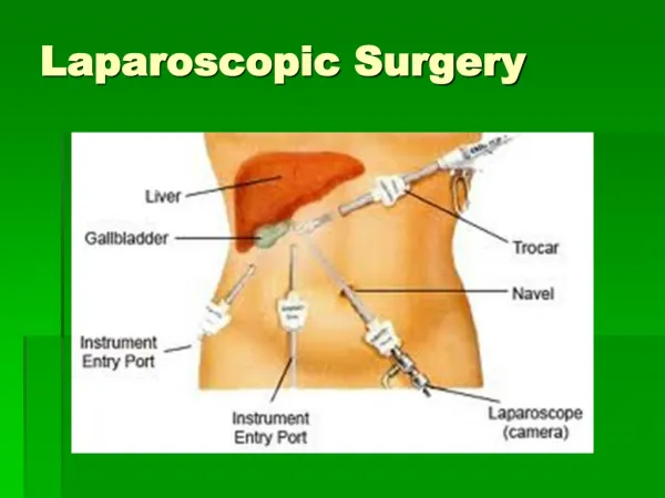
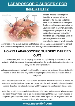
Laparoscopic surfgery • Surgical procedures have been improved to reduce trauma to the patient, morbidity, mortality, and hospital stay, with consequent reductions in health care costs. Starting in the early 1970s, various pathologic gynecologic conditions were diagnosed and treated using laparoscopy. This endoscopic approach was extended to cholecystectomy in the late 1980s. • laparoscopy results in multiple benefits compared with open proceduresand was characterized by better maintenance of homeostasis which explains the effort to use the laparoscopic approach for gastrointestinal (e.g., colonic, gastric, splenic, hepatic surgery), gynecologic (e.g., hysterectomy), urologic (e.g., nephrectomy, prostatectomy), and vascular (e.g., aortic) procedures.
Ventilatory changes • The pneumoperitoneum and the patient positions required for laparoscopy induce pathophysiologic changes that complicate anestheticmanagement. • Pneumoperitoneum decreases thoracopulmonary compliance by 30% to 50%. Reduction in functional residual capacityand development of atelectasis due to elevation of the diaphragmand changes in the distribution of pulmonary ventilation and perfusion from increased airway pressure can be expected.However, increasing IAP to 14 mm Hg with the patient in a 10- to 20-degree head-up or head-down position does not significantly modify either physiologic dead space or shunt in patients without cardiovascular problems. • the partial pressure of arterial carbon dioxide (Paco2) progressively increases to reach a plateau 15 to 30 minutes after the beginning of CO2insufflation.
Any significant increase in Paco2 after this period requires a search for a cause independent of or related to CO2insufflation, such as CO2 subcutaneous emphysema. The increase in Paco2 depends on the IAP.During laparoscopy with local anesthesia, Paco2 remains unchanged but minute ventilation significantly increases. • mean gradients (Δa-ETCO2) between Paco2 and the end-tidal carbon dioxide tension (Petco2) do not change significantly during peritoneal insufflation of CO2. • the lack of correlation between Paco2 and Petco2 in sick patients, particularly those with impaired CO2 excretion capacity. Consequently, hypercapnia can develop, even in the absence of abnormal Petco2. • Postoperative intra-abdominal CO2 retention results in increased respiratory rate and Petco2 of patients breathing spontaneously after laparoscopic cholecystectomy as compared with open cholecystectomy.
the increase of Paco2 may be multifactorial: absorption of CO2 from the peritoneal cavity, impairment of pulmonary ventilation and perfusion by mechanical factors such as abdominal distention, patient position, and volume-controlled mechanical ventilation, the main mechanism being absorption of CO2. • absorption of a gas from the peritoneal cavity depends on its diffusibility, the absorption area, and the perfusion of the walls of that cavity. Because CO2diffusibility is high, absorption of large quantities of CO2 into the blood and the subsequent marked increases in Paco2 would be expected to occur. The limited rise of Paco2 actually observed can be explained by the capacity of the body to store CO2and by impaired local perfusion due to increased IAP.
Respiratory changes during the laparoscopic procedure may contribute to increasing CO2 tension. Mismatched ventilation and pulmonary perfusion can result from the position of the patient and from the increased airway pressures associated with abdominal distention. • At higher IAPs, the continued rise of Paco2 without a corresponding increase in results from an increase in respiratory dead space. If controlled ventilation is not adjusted in response to the increased dead space, alveolar ventilation will decrease and Paco2 will rise. • Paco2 should be maintained within a physiologic range by adjusting the mechanical ventilation. Except in special circumstances, such as when CO2 subcutaneous emphysema occurs, correction of increased Paco2 can be easily achieved by a 10% to 25% increase in alveolar ventilation.
Haemodynamic changes • Multifactorial: result from the combined effects of pneumoperitoneum, patient position, anesthesia, and hypercapnia from the absorbed CO2,reflex increases of vagal tone and arrhythmias. • IN HEALTHY PATIENTS: • IAP higher than 10 mm Hg induces significant alterations of hemodynamics. Results in decrease in cardiac output (10 to 30%), increased arterial pressures, and elevation of systemic and pulmonary vascular resistances. • Heart rates remain unchanged or increased only slightly. The decrease in cardiac output is proportional to the increase in IAP. Cardiac output has also been reported to be increased or unchanged during pneumoperitoneum. • changes in cardiac output are well tolerated by healthy patients.Cardiac outputs, which decrease shortly after the beginning of the peritoneal insufflation, subsequently increase, probably as a result of surgical stress.
mechanism of the decrease of cardiac output is multifactorial: dec venous return (venacavalcompression, pooling of blood in the legs, and an increase in venous resistance) reduction in left ventricular end-diastolic volume Cardiac filling pressures rise during peritoneal insufflation due to increased intrathoracic pressure. Right atrial pressure and pulmonary artery occlusion pressure not reliable indices of cardiac filling pressures during pneumoperitoneum. • Increased filling pressures can be achieved by fluid loading or tilting the patient to a slight head-down position before peritoneal insufflation, by preventing the pooling of blood with intermittent sequential pneumatic compression device,orby wrapping the legs with elastic bandages.
increase in systemic vascular resistance during the existence of the pneumoperitoneum. not a reflex sympathetic response to the decreased cardiac output.Although the normal heart tolerates increases in afterload under physiologic conditions,canbe deleterious to patients with cardiac disease. • The Trendelenburg position attenuates this increase in SVR, the head-up position aggravates it.The increase in systemic vascular resistance can be corrected by the administration of vasodilatinganesthetic agents, such as isoflurane,or direct vasodilating drugs, such as nitroglycerinor nicardipine. • mediated by mechanical and neurohumoralfactors. Catecholamines, the renin-angiotensin system, and especially vasopressin contribute to increasing the afterload.
Increases in plasma vasopressin concentrations correlate with changes in intrathoracic pressure and transmural right atrial pressure. Mechanical stimulation of peritoneal receptors also results in increased vasopressin release,systemic vascular resistance, and arterial pressure. • The increase in SVR also explains why the arterial pressure increases but the cardiac output falls.α2-adrenergic agonists such as clonidineor dexmedetomidineand of β-blocking agents significantly reduces hemodynamic changes and anesthetic requirements. Use of high doses of remifentanil almost completely prevents the hemodynamic changes. • Increased IAP and the head-up position result in lower limb venous stasis. may predispose to the development of thromboemboliccomplications. • Urine output, renal plasma flow, and glomerular filtration rate decrease to less than 50% of baseline values during laparoscopic cholecystectomy.
IN HIGH RISK CARDIAC PATIENTS: In patients with mild to severe cardiac disease, the pattern of change in haemodynamic parameters is qualitatively similar to that in healthy patients but Quantitatively, these changes are more marked. • IV nitroglycerin, nicardipine, or dobutamine has been used in selected patients with heart disease.Nitroglycerinwas chosen to correct the reduction in cardiac output associated with increased pulmonary capillary occlusion pressures and systemic vascular resistance. • nicardipinemay be more appropriate than nitroglycerinas it acts selectively on arterial resistance vessels and does not compromise venous return.Thisdrug is beneficial in case of congestive heart failure which can develop in the early postoperative period Because normalization of hemodynamic variables does not occur for at least 1 hour postoperatively.
CARDIAC ARRHYTHMIAS DURING LAPAROSCOPY: The increased Paco2 may not be the cause of the arrhythmias occurring during laparoscopy. Arrhythmias do not correlate with the level of the Paco2 and may develop early during insufflation, when high Paco2 is not present. • Reflex increases of vagal tone may result from sudden stretching of the peritoneum and during electrocoagulation of the fallopian tubes. Bradycardia, cardiac arrhythmias, and asystole can develop. Vagal stimulation is accentuated if the level of anesthesia is too superficial or if the patient is taking β-blocking drugs. Treatment consists of interruption of insufflation, atropine administration, and deepening of anesthesia. • arrhythmias may also reflect intolerance of the hemodynamic disturbances in patients with known or latent cardiac disease. Gas embolism can also result in cardiac arrhythmias.
Problems due to positioning • the head-down position results in an increase in central venous pressure and cardiac output. The baroreceptorreflex consists of systemic vasodilation and bradycardia. • elevation of the intraocular venous pressure can worsen acute glaucoma. Although the intravascular pressure increases in the upper torso, the head-down position decreases transmural pressures in the pelvic viscera, reducing blood loss but increasing the risk of gas embolism. • With the head-up position, a decrease in cardiac output and mean arterial pressure results from the reduction in venous return.This compounds the hemodynamic changes induced by pneumoperitoneum. The steeper the tilt, the greater the fall in cardiac output. • The head-down position facilitates the development of atelectasis. Steep head-down tilt results in decreases in the functional residual capacity, the total lung volume, and the pulmonary compliance. These changes are more marked in obese, elderly, and debilitated patients.
Post operative benefits • the laparoscopic approach allows for a reduction of the acute phase reaction seen after open cholecystectomy. The metabolic response is also reduced after laparoscopy.It avoids prolonged exposure and manipulation of the intestines and decreases the need for peritoneal incision and trauma. Consequently, postoperative ileus and fasting, duration of intravenous infusion, and hospital stay are significantly reduced after laparoscopy. • Laparoscopy allows a significant reduction in postoperative pain and analgesic consumption. • after laparotomy, patients complain more of parietal pain , whereas after laparoscopic cholecystectomy, patients report also visceral pain, pelvic spasm , and shoulder-tip pain resulting from diaphragmatic irritation. Residual CO2pneumoperitoneum contributes to postoperative pain. • Benefits of intraperitoneal local anesthetic are greater after gynecologiclaparoscopy. Preoperative administration of nonsteroidal anti-inflammatory drugs (NSAIDs) and of cyclooxygenase-2 inhibitors decreases pain.Dexamethasone is also effective in reducing postoperative pain.
Respiratory dysfunction is less severe and recovery is quicker after laparoscopy but diaphragmatic function remains significantly impaired after laparoscopy.Thoracic epidural analgesia does not improve lung function after laparoscopic cholecystectomy. Postoperative pulmonary function of these patients, however, is improved after laparoscopy as compared with laparotomy. • postoperative nausea and vomiting (PONV) (40% to 75% of patients).Whereas perioperativeopioids increase the incidence of PONV,propofolanesthesia can markedly reduce the high incidence of these side effects. • Intraoperativedrainage of gastric contents also reduces PONV.Intraoperativeadministration of droperidol and a 5-hydroxytryptamine type 3 antagonist appears to be helpful in the prevention and treatment of these side effects.Transdermalscopolamine reduces nausea and vomiting after outpatient laparoscopy. • Perioperativeliberal intravenous fluid therapy can contribute to decreasing these symptoms and to improve postoperative recovery.
Alternatives to CO2Pneumoperitoneum • Insufflation of inert gas (e.g., helium, argon) instead of CO2 avoids the increase in Paco2 from absorptionso hyperventilation is not required.Also, the ventilatory consequences of the increased IAP persist. The hemodynamic changes are similar to those observed with CO2. However, the use of these gases accentuates the decrease in cardiac output, whereas the increase in arterial pressure is attenuated. • Unfortunately, the low blood solubility of the inert gases raises the issue of safety in the event of gas embolism. • Another alternative is gasless laparoscopy. The peritoneal cavity is expanded using abdominal wall lift obtained with a fan retractor. This technique avoids the hemodynamic and respiratory repercussions of increased IAP and the consequences of the use of CO2. gasless laparoscopy compromises surgical exposure and increases technical difficulty
complications • CO2 Subcutaneous Emphysema: can develop as a complication of accidental extraperitonealinsufflationbut can also an unavoidable side effect of certain procedures that require intentional extraperitonealinsufflation, such as inguinal hernia repair, renal surgery, and pelvic lymphadenectomy. • Any increase in Petco2 occurring after Petco2 has plateaued should suggest this complication. prevention of hypercapnia by adjustment of ventilation becomes almost impossible. • laparoscopy must be temporarily interrupted to allow CO2 elimination and can be resumed after correction of hypercapnia using a lower insufflationpressure.CO2pressure determines the extent of the emphysema and the magnitude of CO2 absorption. • patient may be mechanically ventilated until hypercapnia is corrected, particularly in COPD patients, to avoid an excessive increase in the work of breathing.
Pneumothorax, Pneumomediastinum, Pneumopericardium: Embryonic remnants constitute potential channels of communication between the peritoneal cavity and the pleural and pericardial sacs, which can open when intraperitoneal pressure increases. Defects in the diaphragm or weak points in the aortic and esophageal hiatus may allow gas passage into the thorax. Pneumothoraces may also develop secondary to pleural tears during laparoscopic surgical procedures at the level of the gastroesophagealjunction. • Capnothorax (CO2 causing a pneumothorax) reduces thoracopulmonary compliance and increases airway pressures. • absorption from the pleural cavity is greater than from the peritoneal cavity; Paco2and Petco2 also increase. • spontaneous resolution of the pneumothorax occurs within 30 to 60 minutes without thoracocentesis.When capnothorax develops during laparoscopy, treatment with positive end-expiratory pressure (PEEP) is an alternative to chest tube placement, but if the pneumothorax is secondary to rupture of preexistingbullae, PEEP must not be applied and thoracocentesis is mandatory.
EndobronchialIntubation: Cephaladdisplacement of the diaphragm during pneumoperitoneum results in cephalad movement of the carina potentially leading to an endobronchialintubation. Generally occurs during procedures in the head-down positionand in the head-up position.results in a decrease in the oxygen saturation with an increase in plateau airway pressure. • Risk of Aspiration of Gastric Contents: Patients undergoing laparoscopy might be considered to be at risk for acid aspiration syndrome . However, the increased IAP results in changes of the lower esophageal sphincter that allow maintenance of the pressure gradient across the gastroesophageal junction and that may therefore reduce the risk of regurgitation. Furthermore, the head-down position should help to prevent any regurgitated fluid from entering the airway, provided airway is secured or airway reflexes are not obtunded.
Gas Embolism: most feared and dangerous complication of laparoscopy. Intravascular injection of gas may follow direct needle or trocar placement into a vessel, or it may occur as a consequence of gas insufflation into an abdominal organ. develops principally during the induction of pneumoperitoneum, particularly in patients with previous abdominal surgery. • CO2 is used most frequently as it is more soluble in blood. Rapid elimination also increases the margin of safety in case of intravenous injection of CO2. this explains the rapid reversal of the clinical signs of CO2 embolism with treatment. Consequently, the lethal dose of embolized CO2 is approximately five times greater than that of air. • Volume preload diminishes the risk of gas embolism and of paradoxical embolism. Ventilation-perfusion mismatching develops with increases in physiologic dead space and hypoxemia.
Early events, occurring with 0.5 mL/kg of air or less, include changes in Doppler sounds and increased mean pulmonary artery pressure. When the size of the embolus increases (2 mL/kg of air), tachycardia, cardiac arrhythmias, hypotension, increased central venous pressure, alteration in heart tones (i.e., millwheel murmur), cyanosis, and ECG changes of right-sided heart strain can develop. • Pulmonary edema can also be an early sign. pulse oximetry, capnometry and capnographyare valuable in providing early diagnosis of gas embolism and determining the extent of the embolism. Petco2 decreases in the case of embolism due to fall in cardiac output and the enlargement of the physiologic dead space. • Initially there may be increase in Petco2 secondary to pulmonary excretion of the CO2, which has been absorbed into the blood. Aspiration of gas or foamy blood from a central venous line is also confirmatory.
Management: immediate cessation of insufflation and release of the pneumoperitoneum. The patient is placed in steep head-down and left lateral decubitus (Durant) position. • Discontinue N2O to allow ventilation with 100% O2 to correct hypoxemia and reduce the size of the gas embolus. • Hyperventilation increases CO2 excretion and is required for increased physiologic dead space. • a central venous or pulmonary artery catheter may be introduced for aspiration of the gas. • Cardiopulmonary resuscitation must be initiated if necessary. External cardiac massage may be helpful in fragmenting CO2 emboli into small bubbles. • The high solubility of CO2 in blood, results in rapid absorption from the bloodstream,andclinical signs of CO2embolism revert rapidly.
Complications of Laparoscopy • Intestinal injuries account for 30% to 50% of these and remain undiagnosed during laparoscopy in one half of the cases. Vascular complications also account for 30% to 50%. Burns were responsible for 15% to 20% of the reported complications. • Bowel perforation, common bile duct injury, and significant hemorrhageare seen in lap cholecystectomy. Laparoscopic cholecystectomy was accompanied by a greater frequency of minor operative complications, whereas open cholecystectomy had a more frequent rate of minor general complications. • retroperitoneal hematoma can develop insidiously and result in significant blood loss without major intraperitoneal effusion, leading to delayed diagnosis. During gynecologic laparoscopy, complications occur more frequently during the creation of pneumoperitoneum and the introduction of trocars, whereas during gastrointestinal surgery they are more closely related to the surgical procedure itself.
Pre operative evaluation • Pneumoperitoneum is undesirable in patients with increased intracranial pressure (e.g., tumor, hydrocephalus, head trauma) and hypovolemia. Laparoscopy can be performed safely in patients with ventricular peritoneal shunt and peritoneojugular shunt that are provided with unidirectional valve resistant to IAPs used during pneumoperitoneum. • In patients with heart disease, cardiac function should be evaluated , particularly in case of compromised ventricular function . Patients with severe congestive heart failure and terminal valvular insufficiency are more prone to develop cardiac complications than patients with ischemic cardiac disease during laparoscopy.
If left ventricular ejection fraction < 30%: pre op echocardiography. Intraoperativemonitoring: Intra-arterial line, Pulmonary artery catheter,Transesophageal echocardiography, Continuous ST-segment analysis. Gasless laparoscopy or laparotomy may be considered. • IntraoperativeManagement: Slow insufflation,Lowintra-abdominal pressure ,Hemodynamic optimization before pneumoperitoneum (preload augmentation) Patient tilt after insufflation. Use ofremifentanil, vasodilatinganesthetic and drugs (nicardipine, nitroglycerin), cardiotonicagents. Preferably an experienced surgeon. • patients with renal failure deserve special care to optimize hemodynamics during pneumoperitoneum, and the concomitant use of nephrotoxic drugs should be avoided.
Anaesthetic technique • General anaesthesia: General anesthesia with endotracheal intubation and controlled ventilation is the safest and most commonly used technique and therefore is recommended for inpatients and for long laparoscopic procedures. • controlled ventilation must be adjusted to maintain Petco2 between 35 and 40 mm Hg;15% to 25% increase of minute ventilation, except when CO2 subcutaneous emphysema develops. Increase of respiratory rate rather than of tidal volume may be preferable in patients with COPD and in patients with a history of spontaneous pneumothorax or bullousemphysema. • The laryngeal mask airway results in fewer cases of sore throat and may be proposed as an alternative to endotrachealintubation;does not protect the airway from aspiration of gastric contents. decreased thoracopulmonary compliance during pneumoperitoneum frequently results in airway pressures exceeding 20 cm H2O. The ProSeal laryngeal mask airway may be an alternative to guarantee an airway seal up to 30 cm H2O. • Local and regional anesthesia