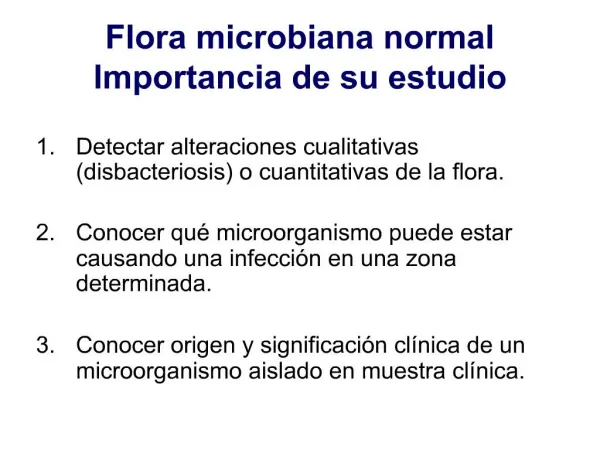
Normal Flora
Normal Flora. Dermatology BM 2023 Lecture 5 Dr Tim Scott-Taylor Health and Human Sciences. Topics. Information covered ; relations to host significance of flora position and density (various areas) bacteria involved benefits of bacterial flora potential problems.

Normal Flora
E N D
Presentation Transcript
Normal Flora • Dermatology BM 2023 • Lecture 5 • Dr Tim Scott-Taylor • Health and Human Sciences
Topics Information covered; • relations to host • significance of flora • position and density (various areas) • bacteria involved • benefits of bacterial flora • potential problems
Learning Objectives • Where normal flora comes from • Details of the prominent bacteria on skin • How they attach and where they reside • benefits of normal flora • Significant examples of opportunistic infections
Normal Flora Present on all exposed skin surfaces Only tissues and certain internal surfaces are sterile Almost exclusively bacteria (protozoa) Usually benign, but potential for opportunistic infection
Surfaces • Internal tissue: eg blood, brain, muscle normally free of microrganisms • Surface tissues: eg skin and mucus membranes constantly in contact environment readily colonised by some species • Internal mucosa: eg urethra, vagina, respiratory tract low density specific organisms The mixture of organisms regularly found at surface anatomical sites is referred to as the normal flora or microbiota
Microbes • Microbes; microscopic; viruses bacteria fungi parasites. • Man is surrounded by microbes. • Most of these are innocuous and are unlikely to pose a major threat to animals. • saprophytes are harmless microbes that live on inanimate material and derive their nutrition from these environmental sources. • symbionts require a human or animal host to survive and multiply. Symbiosis does not distinguish between relationships that are harmful or beneficial found in virtually all environments
Symbiotic Relationships • Mutualism: two organisms from different species living in close proximity to one another and relying on one another for nutrients, protection or other life functions. Both organisms involved benefit from the relationship. • Commensalism: two organisms from different species living in close proximity to one another, in which one member is unaffected by the relationship and the other benefits from it • Parasitism: two organisms from different species living in close proximity to one another, in which one member depends on another for its nutrients, protection or other life functions. The parasite benefits from the relationship while the host is harmed by it • Amensalism: two organisms from different species living in close proximity to one another, where one of the members suffers as a result of the relationship while the other is unaffected by it
Commensalism • normal flora are commensals that derive food and shelter from the host. They normally do no harm to the host. The host may benefit from the presence of the microbe. • relationship also described as mutualism. No harm is done to the host. • Commensal microbes colonise the host • Infection implies that harm is done to the host i.e. causes disease. A microbe that causes disease is a pathogen. • Usually the host will manifest an inflammatory response to a pathogen, but not to a coloniser at a normally non-sterile body site
Pathogen • a microbe that can initiate infection, often with only small numbers • strict pathogens i.e. will always cause disease eg Bacillus anthracis (anthrax). • Some pathogens may sometimes almost behave like commensals, eg Salmonella typhi (typhoid) (carrier state). • Whether a microbe behaves as a pathogen i.e. causes disease, depends on the properties of the microbe and the host. • There can be a fine balance between microbial and host properties; ImmunityVirulence Resistance Disease microbiota
Nature of Normal Flora • A few Archaebacteria in the gut: Methanobrevibacter smithii • Some prokaryotes, Candida albicans in mouth Pneumocystis carinii in pharynx Entamoeba gingivalis between teeth
Tooth Amoebas • Entamoeba gingivalis • microscopic parasites 10-35 μm • Live in crevices between teeth and gums. Brushing does not remove them • Beneficial; eat mouth bacteria only harmful when v. numerous lack of oral hygiene • No cysts are formed; transmission is entirely by oral-oral contact.
Nature of Normal Flora • A few Archeabacteria in the gut • Some prokaryotes, Candida albicans in mouth Pneumocystis carinii in pharynx Entamoeba gingivalis • But microbiota almost exclusively bacterial
Numbers • Human body = ~1013 cells • Bacteria; 1010 in the mouth 1012 on the skin1014 in intestine • Skin low density 100-1000s/cm2 ; except axillae groin toe • Density varies; age sex diet nutrition Far in excess of number of eukaryote cells in all organs high moisture level
Bacterial Staining Bacteria distinguished primarily on shape; cocci / bacilli staining; gram +ve / -ve Gram stain Divides bacteria into Gram positive – blue/purple Gram negative- red Hans Christian Gram, Danish bacteriologist devised stain in 1882
Gram Staining Gram negative Gram positive
Gram’s Stain Procedure • Place a slide with a bacterial smear on a staining rack. • Stain the slide with crystal violet for 1-2 min. • Pour off the stain. • Flood slide with Gram's iodine for 1-2 min. • Pour off the iodine. • Decolourize the slide briefly with acetone (2-3 seconds). • Wash slide thoroughly with water to remove the acetone • Flood slide with safranin counterstain for 2 min. • Wash with water. • Blot excess water and dry in hand over bunsen flame.
How Does it Work? • Gram didn't know - he simply worked empirically • Gram reaction is based on the structure of the bacterial cell wall • In Gram-positive bacteria, the purple crystal violet stain is trapped by the layer of peptidoglycan which forms the outer layer of the cell • In Gram-negative bacteria, the outer membrane prevents the stain from reaching the peptidoglycan layer in the periplasm. The outer membrane is then permeabilized by acetone treatment, and the pink safranin counterstain is trapped by the peptidoglycan layer.
Bacterial Cell Walls
Four Basic Types Gram - cocci Gram + cocci e.g. Staphylococcus aureus e.g. Neisseria Gram + rod Gram - rod e.g. Bacillus cereus e.g. Escherichia coli
Common Bacteria in Flora ++ = nearly 100 percent + = common +/- = rare * = potential pathogen
Skin • Not a great habitat; dries out, constantly being shed, secretions include fatty acids (lower pH to 4-6) and salt. • Some skin regions better habitats than others: scalp, ears, underarms, anal region are all especially good • Bacteria that can grow on skin must be able to survive these conditions. • Typical bacteria: Staphylococcus epidermidis, not normally a pathogen: but infection via surgical implants and catheters • Propionobacterium acnes; lives in sweat glands, hair follicles, not eliminated by washing skin.
Staphylococci Gram positive cocci in clusters • Greek staphyle = bunch of grapes • S. aureus : cause of soft tissue infections toxic shock syndrome (TSS) scalded skin syndrome. • S. epidermidis : common on skin, coagulase-negative coagulase test clot plasma
Corynebacteria Gram positive rod (bacillus) Pallisades Chinese writing • common on skin and in GI tract • irregular pleomorphic with metachromatic granules • grey / black colonies on tellurite medium • C. diphtheriae throat pathogen potent toxin
Streptococci Gram positive cocci in chains • Streptococci occur on all skin and mucosal surfaces • glycocalyx: adherence to plastic and cells resistance to phagocytosis and antibiotics. • Streptococcus pyogenes common on skin but causes erysipelas, scarlet fever, rheumatic fever
Conjunctiva • Not completely sterile, numbers of bacteria is small Lachrymal secretions contain lysozyme Blinking continually wipes away bacteria • Staphylococcus epidermidis Corynebacteria Blepheritis Conjunctivitis Specific attachment to receptors Haemophilus sialic acid residues Chlamydia dominant
Respiratory Tract Nares (nostrils) are heavily colonised > 200 species Staphylococcus epidermidis Corynebacteria Staphylococcus aureus (20% of population) Upper respiratory tract also highly colonised non-haemolytic Streptococci Neisseria Streptococcus pneumoniae Haemophilus influenzae Lower respiratory tract Mucus tissue damage action of cilia disease dominant most non-pathogenic but some pathogens
Urogenital Tract • Upper urogenital tract; normally sterile action of urine • Anterior urogenital tract;Staphylcoccus epidermidis Escherichia coli • Vagina; Corynebacteria Staphylococci Streptococci Lactobacillus acidophilus: at puberty lactic acid prevents establishment other bacteria Colonise soon after birth
Mouth • Favourable habitat; nutrients and secretions epithelial debris saliva 104-109/ml organisms • Streptococcus salivarious ; 98% predominant until teeth erupt • Flora increasingly complex, anerobes, Bacteroides Streptococcus mutans Streptococcus sanguis Streptococcus pyogenes rheumatic fever damaged heart valves lysozyme Dominant, caries
Dental Caries • Initiated by normal flora, Streptococcus mutans • Dental plaque; consists 60-70% cells salivary polymers bacterial extracelular products • Initiated by glucosyl transferase, surface enzyme initial attachment to tooth via salivary glycoprotein creation of glycan biofilm = bacterial capsule lactic acid from dietary sugars demineralises enamel Lactobacilli secondary Actinomyces israelli proteolytic invaders
Normal Gut Flora relatively few bacteria in anterior bowel increasing diversity and density of bacteria in posterior bowel
Escherichia coli Gram negative rods (bacilli) • type faecal bacterium (enterococus in US) • opportunistic pathogen in urogenital canal • E. coli 0157: haemorrhagic colitis, uncooked meat
GI Tract • digestive enzymes and stomach acid kill bacteria • small intestine has few bacteria, colon huge population 1/3 of faeces is bacteria up to 1000 organisms/gram over 300 different species • E. coli is only 0.1% of total population anaerobic Bacteroides most abundant; ~ 25% microbiota • bacteria in colon divide every 12-24 hours on average, much slower than laboratory batch culture rates.
GI Tract • high flow rates make small intestine difficult to colonize, concentration of bacteria remains low, ~106/ml • takes food ~3-5 hours to move through small intestine takes food 24-48 hours to travel through the colon • slow flow rates allow bacterial multiplication, 1012-1013/ml • 30-50% of contents, ~2-3 lbs weight = bacteria • bacteria breakdown complex polysaccharide; xylan cellulose pectin • colon an organ of digestion where normal flora does most of the work called colonic fermentation
Tissue Tropism Normal flora exhibit a tissue predilection for colonisation Could be due to; - supply of a specific essential growth factor - construction of a biofilm some bacteria are able to colonise the biofilms of others, most biuofilms are a mixture of bacteria - a specific receptor present at some sites
Examples of Tissue Tropism For some of bacteria in normal flora the attachment factors are precisely known
Gnotobiotic Animals • Germfree, axenic; to study effect of normal flora • easy to produce germfree birds; sterilize shell, use sterile incubator • Germfree mammals; cesarean section isolation chambers • Germfree animals are less healthy than animals with normal flora; • Greater vitamin requirements for K and B complex • lower cardiac output • much more susceptible to pathogens • much smaller infectious dose required to initiate an infection • Live shorter lives air, food, water sterilized
Benefits of Normal Flora • Synthesise and secrete vitamins; vitamin K germ-free deficient vitamin B12 • Prevent colonisation by pathogens oral cavity, skin germ free 10 salmonella vs 106gut lactobacilli maintain high pH vagina • Antagonise other bacteria; fatty acids inhibit / kill peroxide bacteriocins • Stimulate certain tissues; Peyer’s patches poorly developed immune system caecum • Stimulate cross-reactive antibody immunise against pathogens
Disadvantages of Normal Flora 1. Body odour • body odour originates from the skin • decoposition of secretions of apocrine sweat glands located primarily under arms and in the groin • Corynebacterium tenuis and C. xerosis in particular • best eliminated through good hygiene • fungal infections such as athlete's foot also odourous
Antibiotic Resistance • MRSA: Methicillin Resistant Staphylococcus aureus • resistant to all commercially available antibiotics, including methicillin and vancomycin • carried in the noses of health care workers and transmitted from patient to patient • major cause of surgical wounds and systemic infections • antibiotic resistance is transferred to other organisms • recent NHS directive to alcohol wash hands between beds has cut incidence by 50%
Opportunistic Infection Some commensals can act as pathogens when host’s defences are weakened or immunocompromised • infection caused by a normally benign commensal eg infection with Candida albicans(yeast) a normal commensal of mouth and gut in immunosuppressed leukaemia patients eg infection with Pneumocystis carinii a low virulence fungus during mmunosuppresion due to HIV
Opportunistic Infection Some organisms that are commensals at one body site may be pathogens at another body site eg Staphylococcus aureus in the nose (commensal) in a post-operative wound infection (pathogen) egEscherichia coli in GI tract (commensal) in urinary tract causes UTI (pathogen).
Dental Caries • Teeth in skulls from Europeans prior to the 1500's showed remarkably well-preserved teeth. • Once sucrose, a dissacharide from cane sugar, was introduced tooth decay became widespread • S. mutans produces a thick capsule of dextran. The gooey polysaccharide forms a biofilm and allows other bacteria to attach Dental Plaque accumulates • S. mutans uses lactic acid fermentation exclusively as its catabolic pathway. Acids attack tooth enamel
Ulcers • Stomach acid attacks duodenal lining; ulcers related to stress; • 1980s Warren and Marshall discovered antibiotics reduced ulceration, awarded Nobel Prize Medicine 2005 • 30-50% of human population carry Helicobacter pylori • Spiral shape and flagella helps bacterium burrow into protective mucous lining. Enzyme urease converts urea into ammonia and bicarbonate, neutralizes stomach acid • ammonia protease catalase phospholipases toxic to duodenal epithelial cells
H. pylori and Gastric cancer • bacterium categorized as group I carcinogen by the International Agency for Research on Cancer (IARC) • Gastric carcinoma MALT lymphoma (mucosa -associated lymphoid tissue associated with H. pylori • Two related mechanisms proposed; 1. free radicals production increases rate of cell mutation 2. TNFα, interleukin 6, inflammation alter cell adhesion proteins and mutate tumor suppressor genes
Summary • Bacteria perform physiological, nutritional and protective functions in the human body. • Maintaining a balance is crucial Flora consists of ecosystems consequences of disruption unpredictable. • normal flora is complexity and understanding of function limited eg < 1% of bacteria grow on laboratory media > 99% the microbial world unexplored antibiotics tissue damage medical procedures changes in diet